Copyright
©The Author(s) 2023.
World J Clin Cases. Oct 16, 2023; 11(29): 7193-7199
Published online Oct 16, 2023. doi: 10.12998/wjcc.v11.i29.7193
Published online Oct 16, 2023. doi: 10.12998/wjcc.v11.i29.7193
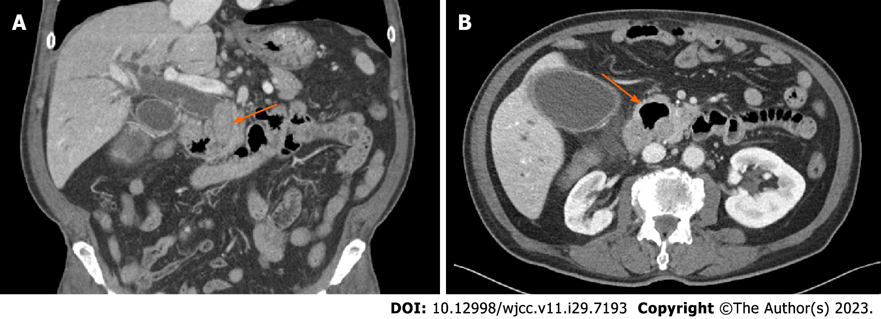
Figure 1 Preoperative computed tomography finding.
A: Impacted large common bile duct stone (arrow); B: Large duodenal diverticulum (arrow).
Figure 2 Location of trocars.
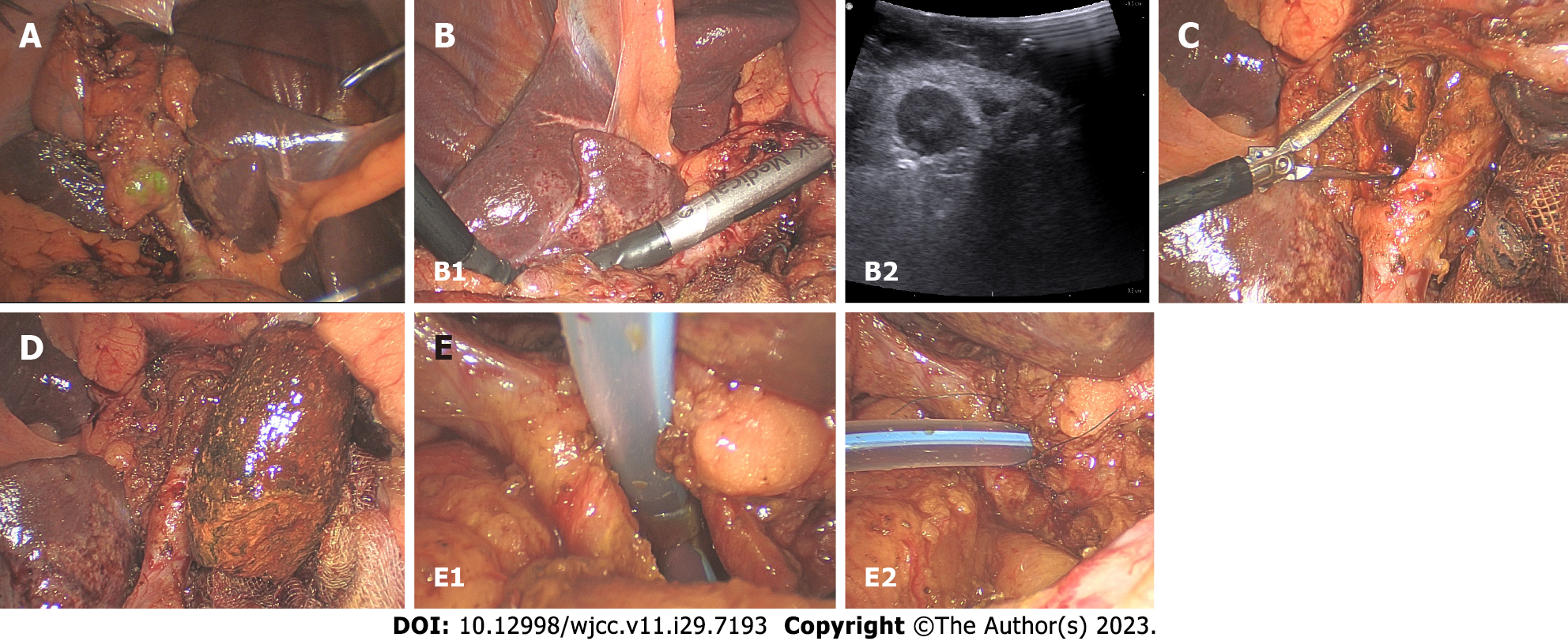
Figure 3 Intraoperative photos.
A: Fixation of the gallbladder on the anterior abdominal wall; B: Intraoperative ultrasound; C: Common bile duct stone removal with the Endo BabcockTM; D: Removed common bile duct stone; E: Transductal T-tube insertion and suturing (left: T-tube insertion; right: after suturing).
- Citation: Yoo D. Laparoscopic choledocholithotomy and transductal T-tube insertion with indocyanine green fluorescence imaging and laparoscopic ultrasound: A case report. World J Clin Cases 2023; 11(29): 7193-7199
- URL: https://www.wjgnet.com/2307-8960/full/v11/i29/7193.htm
- DOI: https://dx.doi.org/10.12998/wjcc.v11.i29.7193


