Copyright
©The Author(s) 2023.
World J Clin Cases. Oct 16, 2023; 11(29): 7170-7178
Published online Oct 16, 2023. doi: 10.12998/wjcc.v11.i29.7170
Published online Oct 16, 2023. doi: 10.12998/wjcc.v11.i29.7170
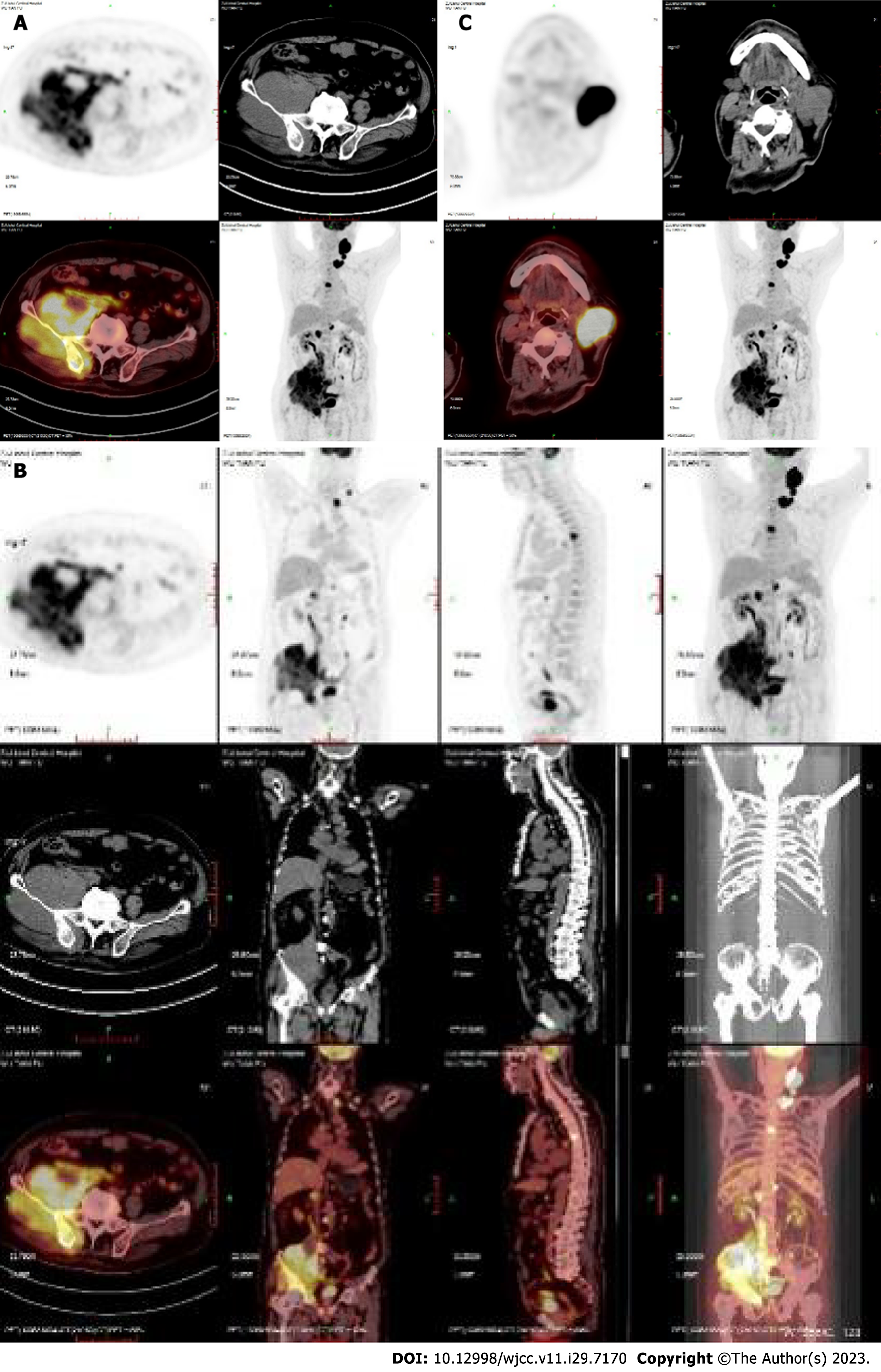
Figure 1 Positron emission tomography-computed tomography scan showed multiple enlarged lymph nodes in the left neck and left supraclavicular region.
The bone density of the right ilium increased unevenly, with an irregular soft tissue mass involving the right side of the sacrum and the right acetabulum, with unclear borders (approximately 12.8 cm × 11.3 cm). A: The iliac bone scan; B: The whole-body scan; C: The neck scan.
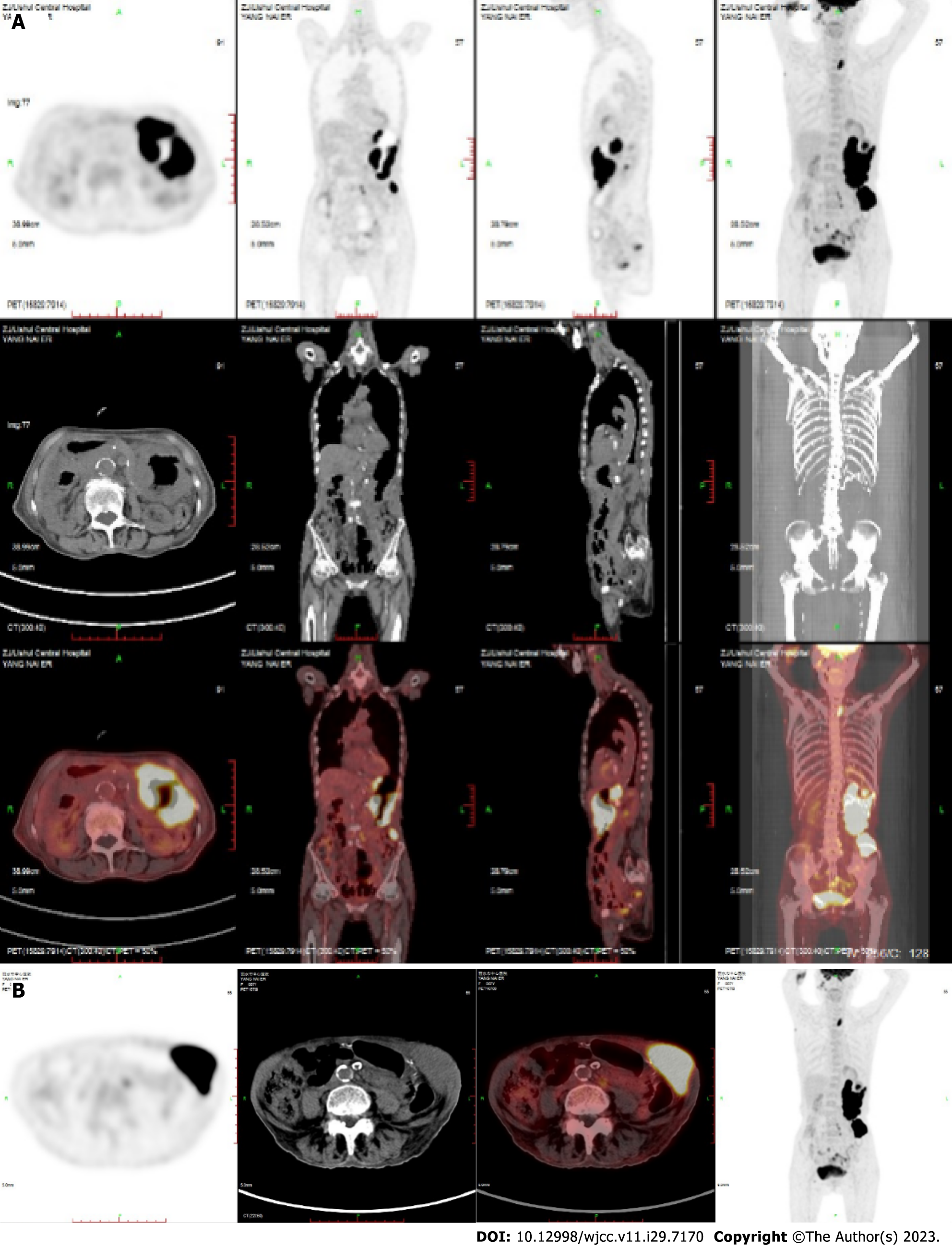
Figure 2 Positron emission tomography-computed tomography showed extensive irregular thickening of the stomach wall from the cardia, and the stomach body was accompanied by multiple ulcers.
A: The whole-body scan; B: The stomach scan.
Figure 3
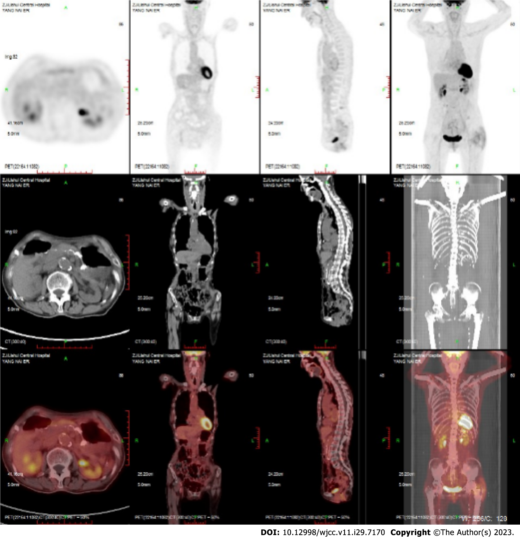
After treatment, positron emission tomography-computed tomography showed complete remission of lymphoma.
Figure 4
Positron emission tomography-computed tomography showed complete remission of lymphoma.
- Citation: Zhang CJ, Zhao ML. Rituximab combined with Bruton tyrosine kinase inhibitor to treat elderly diffuse large B-cell lymphoma patients: Two case reports. World J Clin Cases 2023; 11(29): 7170-7178
- URL: https://www.wjgnet.com/2307-8960/full/v11/i29/7170.htm
- DOI: https://dx.doi.org/10.12998/wjcc.v11.i29.7170


