Copyright
©The Author(s) 2023.
World J Clin Cases. Oct 16, 2023; 11(29): 7150-7155
Published online Oct 16, 2023. doi: 10.12998/wjcc.v11.i29.7150
Published online Oct 16, 2023. doi: 10.12998/wjcc.v11.i29.7150
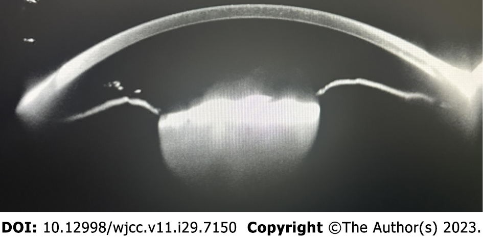
Figure 1
The lens was a dense mass with strong reflection, and the front surface of the lens was uneven, locally “hill-like”, and the rear surface echoes were indistinct.
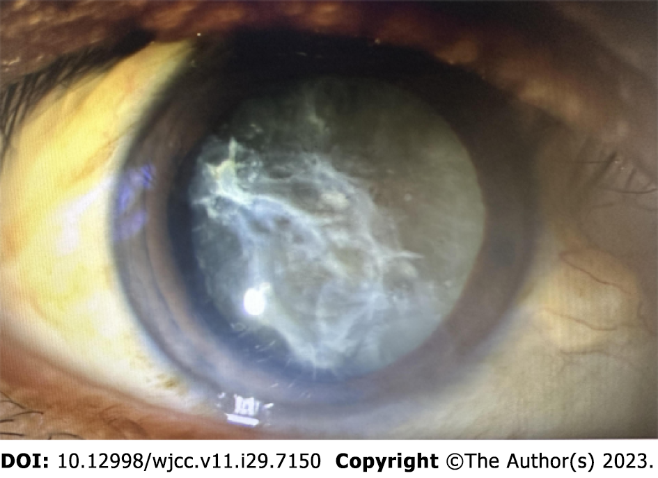
Figure 2 The anterior capsule of the lens was obviously organized.
The density was formed 10 to 4 points organized membrane, a 6-point organic membrane bulge was seen, and the lower capsule was pulled in the direction of the cornea. It was “hilly”, the lens was densely white and cloudy, and the nucleus was grade IV.
Figure 3 The operation process.
A: It was difficult to remove the anterior capsule by mechanization, and a sticky bomb was injected under the anterior capsule to provoke it; B: The 10-point mechanized cord was cut homeopathically; C: Tearing capsule pinch forceps were used to continue tearing the capsule; D: The 8-point mechanized cable was cut; E: Tearing of the capsule continued; F: The 4-point mechanized cable was cut; G: Tearing the capsule continued, and maintain the integrity of the lens capsule without tearing; H: The phacoemulsification needle was placed diagonally downward; I: The interception-splitting method was used to split the lens nucleus into several pieces, and the fragmented nucleus was removed by emulsification in stages.
Figure 4
Intraocular lens implantation in the capsule.
- Citation: Wang LW, Fang SF. Surgical treatment of severe anterior capsular organized hard core cataract: A case report. World J Clin Cases 2023; 11(29): 7150-7155
- URL: https://www.wjgnet.com/2307-8960/full/v11/i29/7150.htm
- DOI: https://dx.doi.org/10.12998/wjcc.v11.i29.7150


