Copyright
©The Author(s) 2023.
World J Clin Cases. Oct 6, 2023; 11(28): 6967-6973
Published online Oct 6, 2023. doi: 10.12998/wjcc.v11.i28.6967
Published online Oct 6, 2023. doi: 10.12998/wjcc.v11.i28.6967
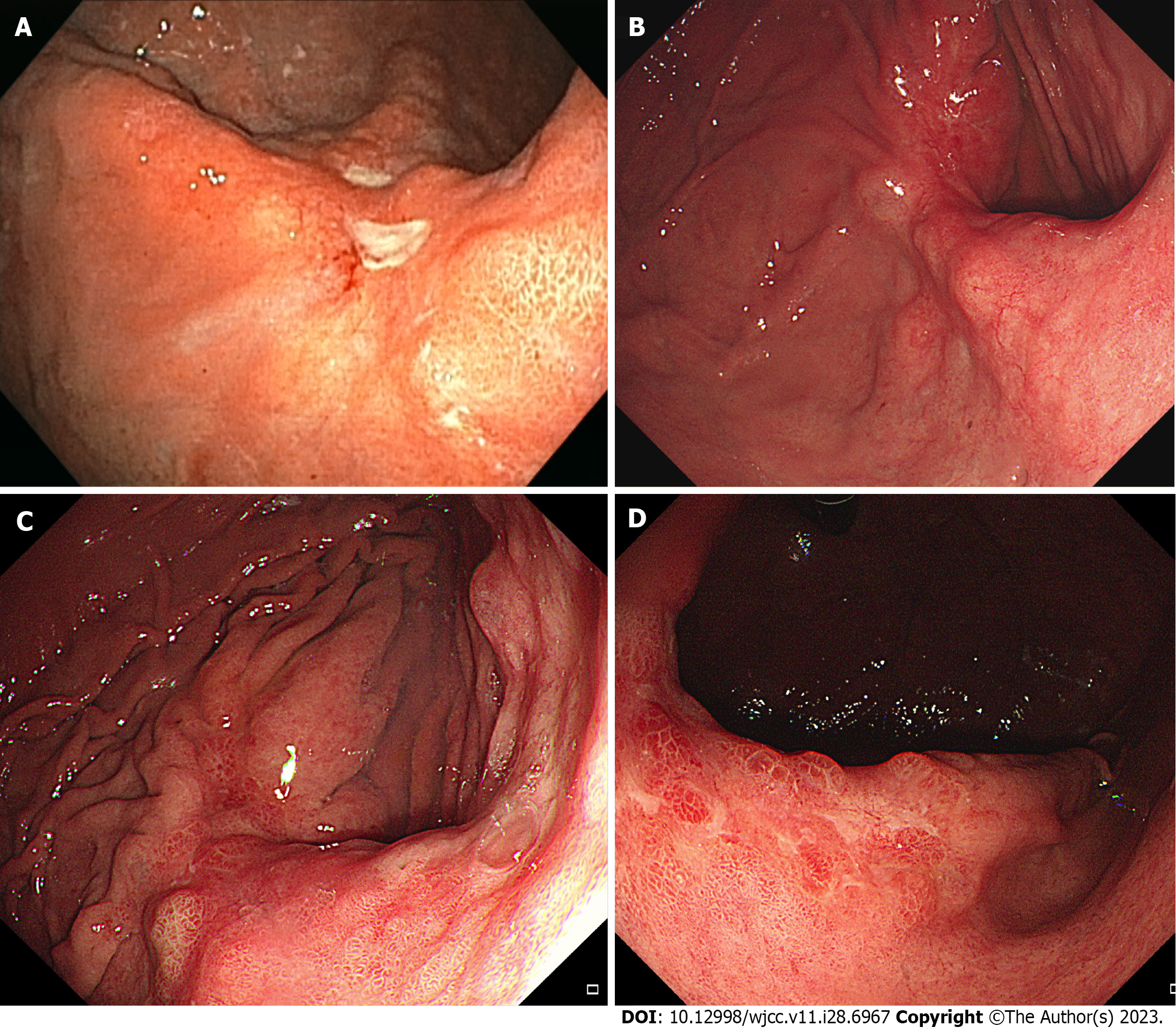
Figure 1 Initial and follow-up endoscopic findings.
A: Initial esophagogastroduodenoscopy (EGD) showing two whitish ulcerative lesions at the posterior wall of the mid-body; B: In the first follow-up EGD, the lesions are healed with a reddish scar and several bulging areas of normal mucosa on the peripheral; C: The last follow-up EGD presents a 4.5 cm-sized ulcero-infiltrative lesion in the posterior wall of the mid-body; D: In the retroflexed view, this lesion is covered with an unevenly distributed regenerative epithelium and whitish discoloration. The tumor is identified as a well-differentiated adenocarcinoma.
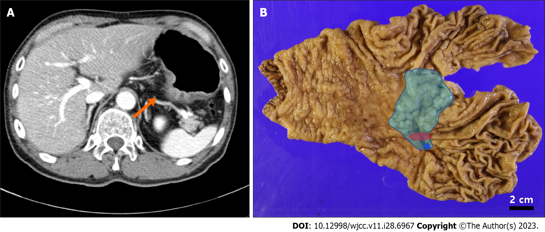
Figure 2 Preoperative image study and gross lesions of the stomach.
A: Preoperative abdominal computed tomography detected a 4.6 cm-sized lesion at the posterior wall of the mid-body (orange arrow); B: On the total gastrectomy specimen, a 6.9 cm × 4.5 cm-sized ulcero-infiltrative gastric hamartomatous inverted polyps extending from the lesser curvature of the cardia to the posterior wall of the body (blue) and a 1.6 cm-sized adenocarcinoma located at the posterior wall of the body (red) were indicated. The two lesions showed an overlap.
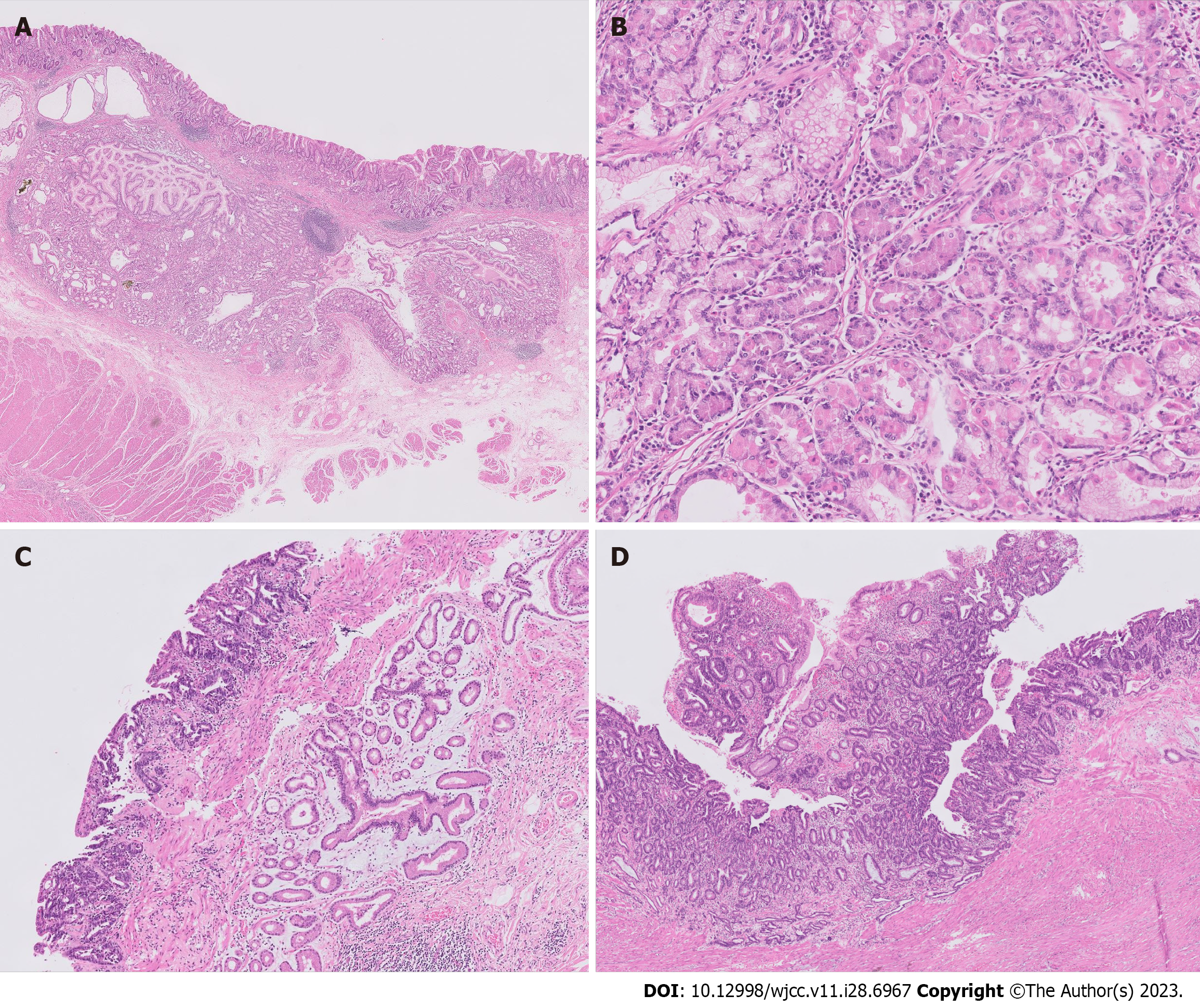
Figure 3 Microscopic findings of the surgical specimen (hematoxylin and eosin staining).
A: Gastric hamartomatous inverted polyp (GHIP) is located in the submucosal layer with focal cystic dilations and is surrounded by smooth muscle bundles. On the left upper side, gastritis cystica profundas is adjacent to the GHIP (20 × magnification); B: GHIP consists of heterogeneous glandular cell types (100 × magnification); C: GHIP is positioned separately under a well differentiated adenocarcinoma (40 × magnification); D: Adenocarcinoma extending to the muscularis propria is shown (40 × magnification).
- Citation: Park G, Kim J, Lee SH, Kim Y. Large gastric hamartomatous inverted polyp accompanied by advanced gastric cancer: A case report. World J Clin Cases 2023; 11(28): 6967-6973
- URL: https://www.wjgnet.com/2307-8960/full/v11/i28/6967.htm
- DOI: https://dx.doi.org/10.12998/wjcc.v11.i28.6967


