Copyright
©The Author(s) 2023.
World J Clin Cases. Oct 6, 2023; 11(28): 6961-6966
Published online Oct 6, 2023. doi: 10.12998/wjcc.v11.i28.6961
Published online Oct 6, 2023. doi: 10.12998/wjcc.v11.i28.6961
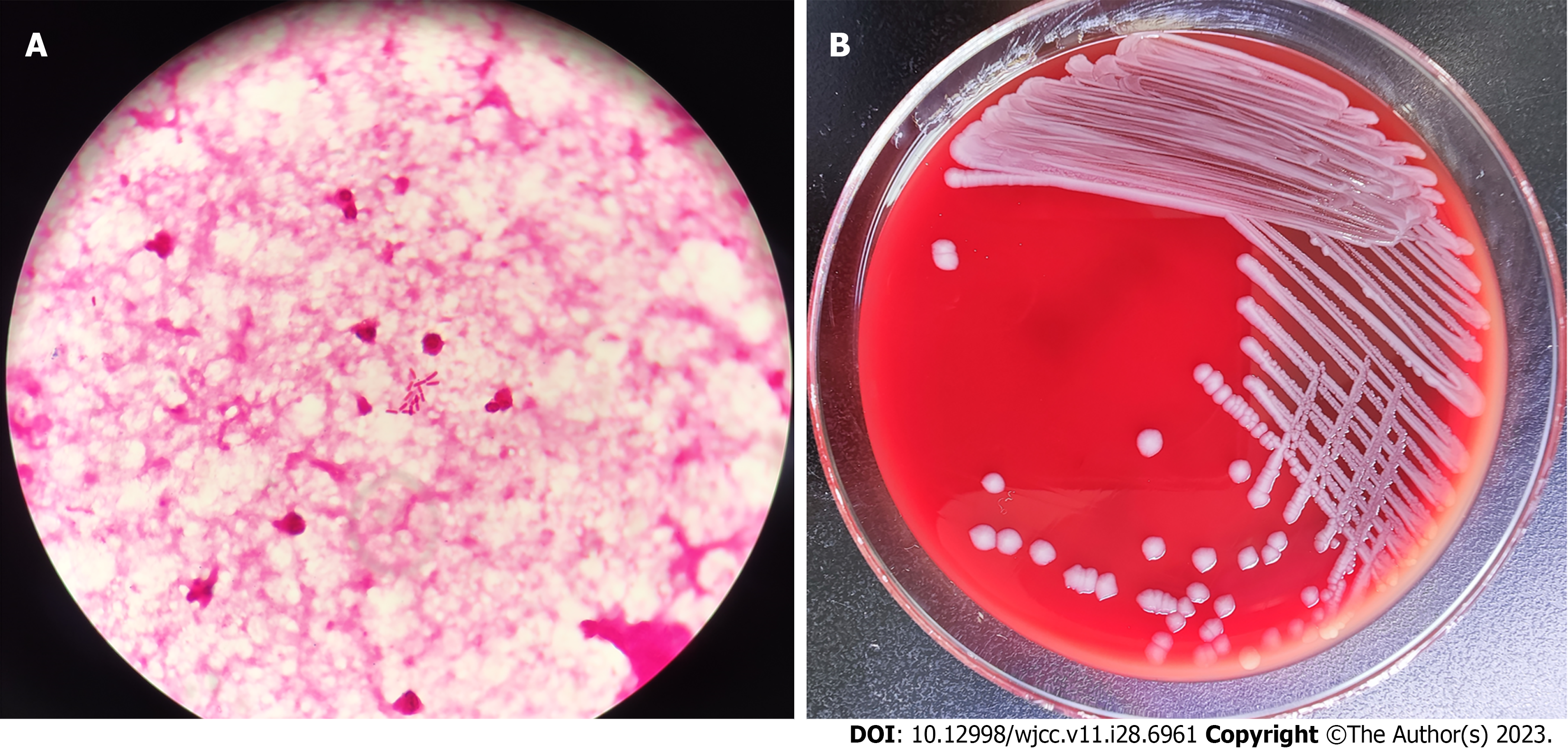
Figure 1 Smear examination and bacterial culture results of cerebrospinal fluid.
A: Gram staining of peritoneal dialysate specimens after centrifugation (1000 ×); B: Colony morphology on a blood agar plate at 35°C, 5% CO2 and cultured for 48 h.
Figure 2 Head magnetic resonance imaging before treatment.
A: Head magnetic resonance imaging (MRI) were observed subacute cerebral infarction; B: MRI suggested hematocele in the posterior horn of bilateral ventricles; C: MRI revealed widening of extracerebral space. MRI: Magnetic resonance imaging.
- Citation: Yu JL, Jiang LL, Dong R, Liu SY. Intracranial infection and sepsis in infants caused by Salmonella derby: A case report. World J Clin Cases 2023; 11(28): 6961-6966
- URL: https://www.wjgnet.com/2307-8960/full/v11/i28/6961.htm
- DOI: https://dx.doi.org/10.12998/wjcc.v11.i28.6961


