Copyright
©The Author(s) 2023.
World J Clin Cases. Oct 6, 2023; 11(28): 6955-6960
Published online Oct 6, 2023. doi: 10.12998/wjcc.v11.i28.6955
Published online Oct 6, 2023. doi: 10.12998/wjcc.v11.i28.6955
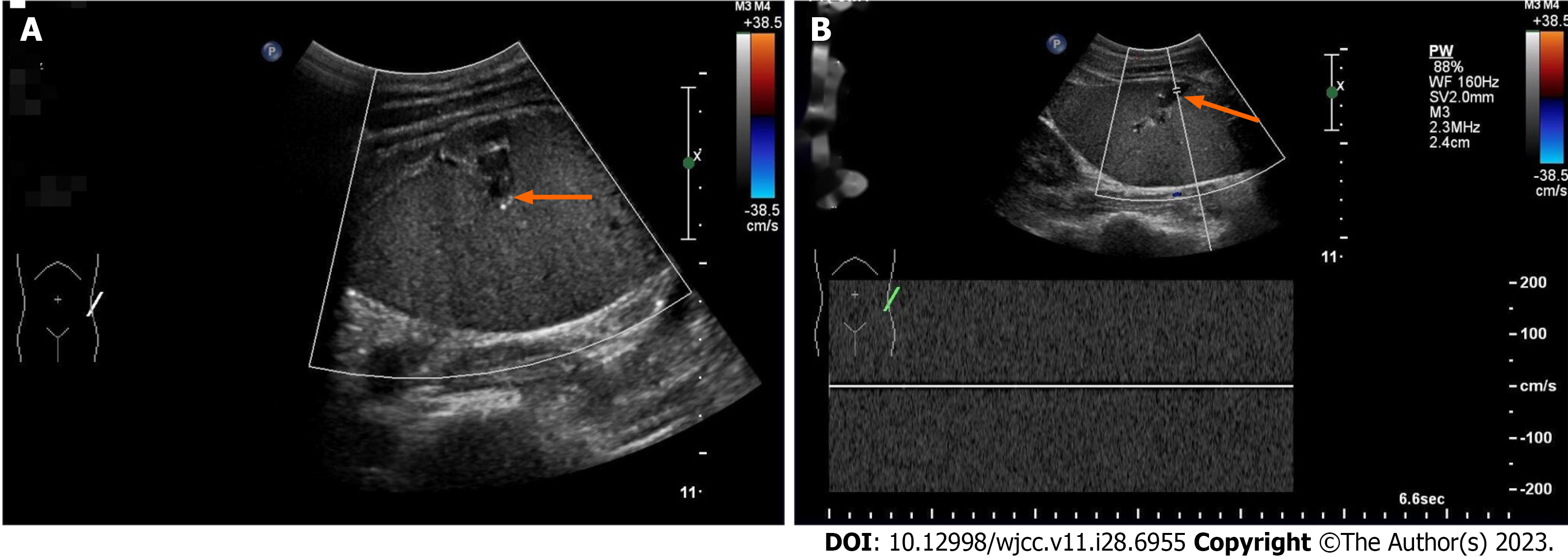
Figure 1 Ultrasound of the abdomen with arrows showing splenic vein thrombosis and hypovascularized spleen.
A: Splenic vein widened without blood flow (orange arrow); B: No venous spectrum can be extracted from splenic vein (orange arrow).
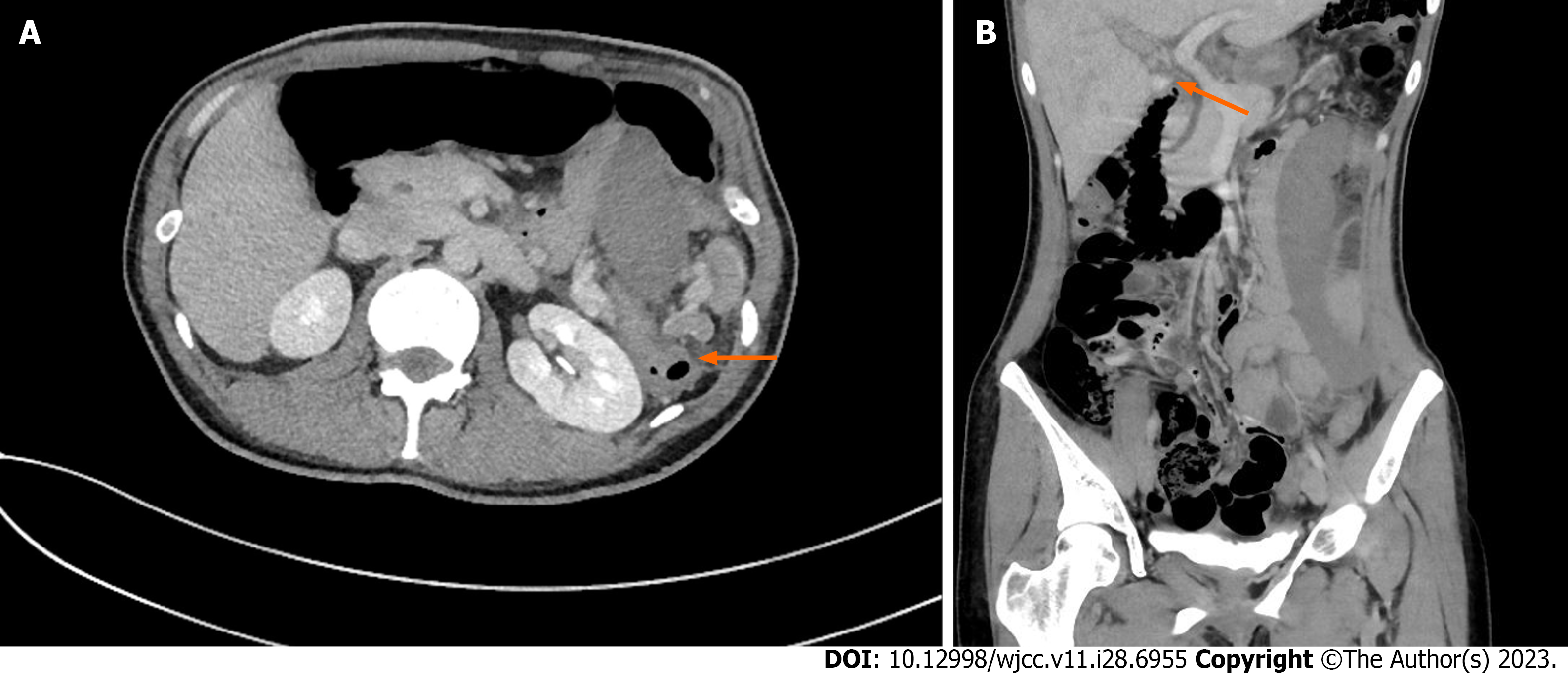
Figure 2 Computed tomography with arrows showing splenic pedicle vascular torsion and portal vein thrombosis.
A: Axial reconstruction planes (orange arrow); B: Coronal reconstruction planes (orange arrow).
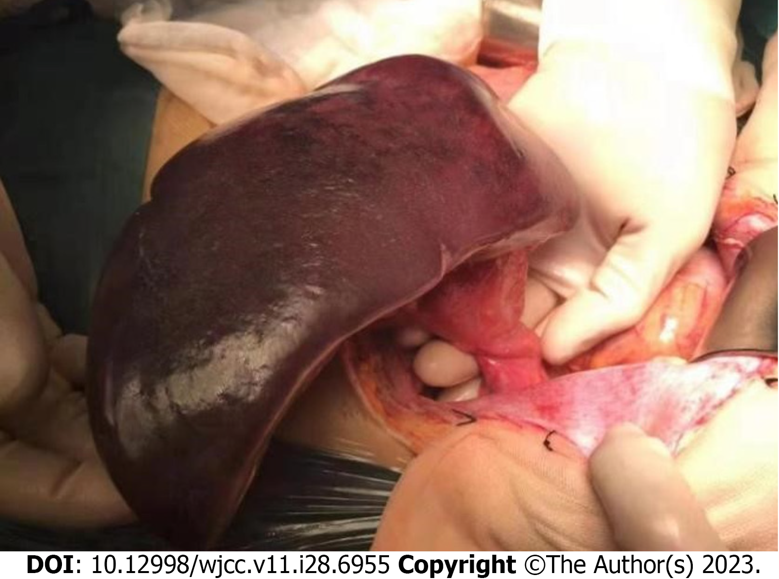
Figure 3 Splenic infarction caused by torsion of splenic pedicle vessels.
- Citation: Zhu XY, Ji DX, Shi WZ, Fu YW, Zhang DK. Wandering spleen torsion with portal vein thrombosis: A case report. World J Clin Cases 2023; 11(28): 6955-6960
- URL: https://www.wjgnet.com/2307-8960/full/v11/i28/6955.htm
- DOI: https://dx.doi.org/10.12998/wjcc.v11.i28.6955


