Copyright
©The Author(s) 2023.
World J Clin Cases. Oct 6, 2023; 11(28): 6949-6954
Published online Oct 6, 2023. doi: 10.12998/wjcc.v11.i28.6949
Published online Oct 6, 2023. doi: 10.12998/wjcc.v11.i28.6949
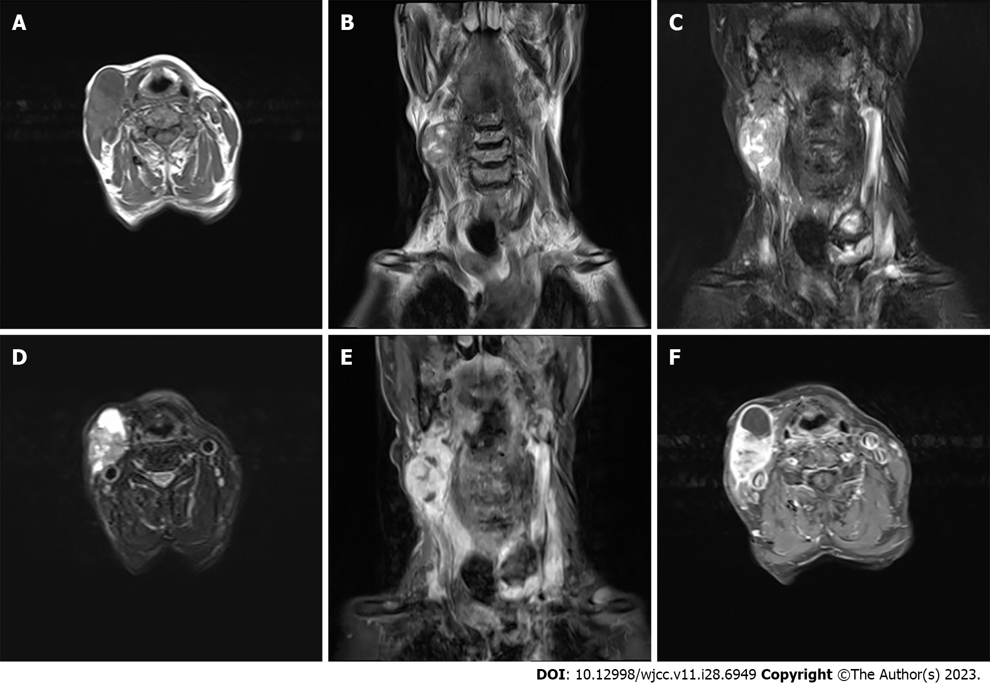
Figure 1 Magnetic resonance imaging scans.
A: Hypointense lesion on T1-weighted images; B and C: Predominantly hypointense and hyperintense lesion on T2-weighted images; D: Mixed signal mass on T2 compression lipid sequence, primarily isointense to hypointense; E and F: Notable heterogeneous enhancement of the mass on contrast-enhanced magnetic resonance imaging.
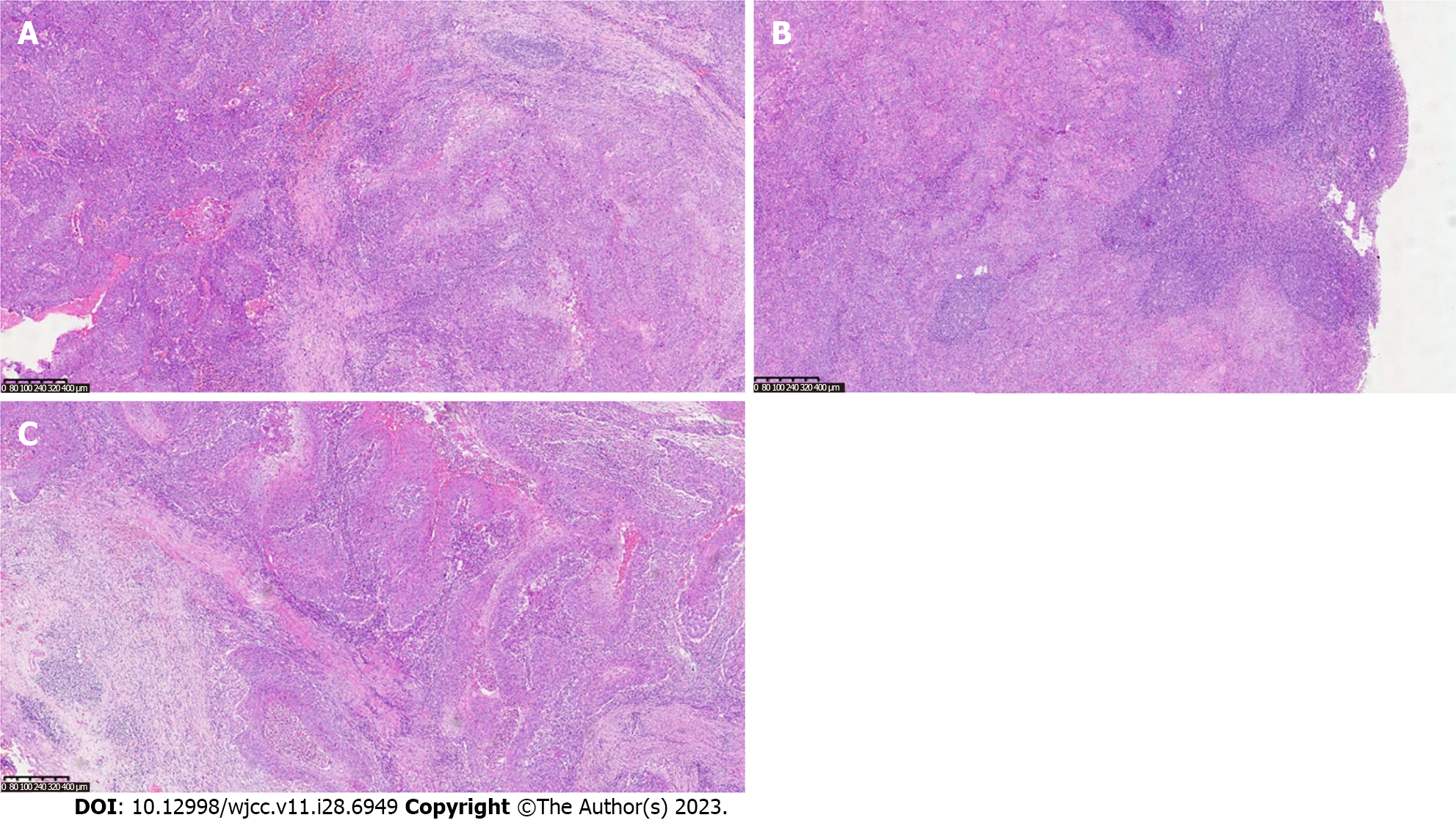
Figure 2 Pathological findings.
A: Vigorous tumor growth [hepatic encephalopathy (HE) × 100)]; B: Lymph node metastasis; C: Tumor nests (HE × 50).
- Citation: Wang K, Wen JZ, Zhou SX, Ye LF, Fang C, Chen Y, Wang HX, Luo X. Malignant proliferative ependymoma of the neck with lymph node metastasis: A case report. World J Clin Cases 2023; 11(28): 6949-6954
- URL: https://www.wjgnet.com/2307-8960/full/v11/i28/6949.htm
- DOI: https://dx.doi.org/10.12998/wjcc.v11.i28.6949


