Copyright
©The Author(s) 2023.
World J Clin Cases. Oct 6, 2023; 11(28): 6943-6948
Published online Oct 6, 2023. doi: 10.12998/wjcc.v11.i28.6943
Published online Oct 6, 2023. doi: 10.12998/wjcc.v11.i28.6943
Figure 1 First abdominopelvic computed tomography results.
A: Axial image; B: Coronal image. Both images show the pre- and peri-vesical areas along with pneumatosis intestinalis near the pelvic small bowel loop.
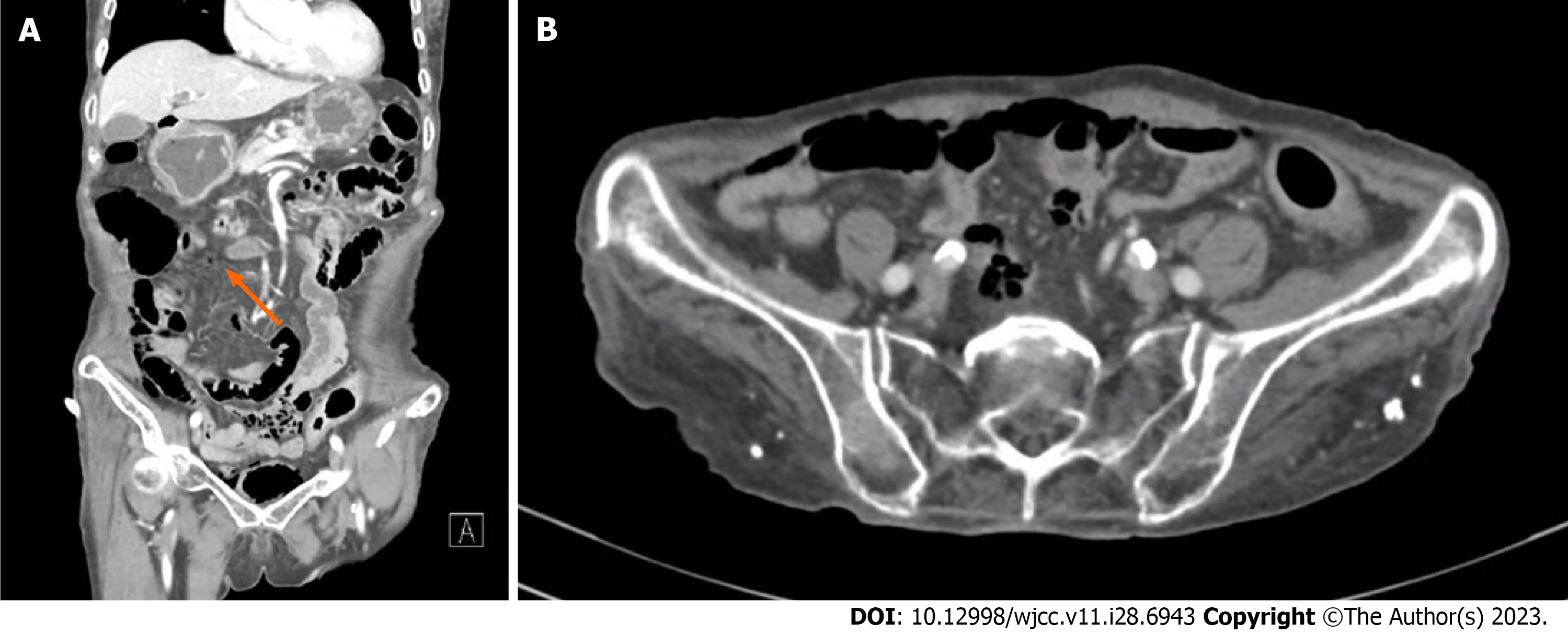
Figure 2 Second abdominopelvic computed tomography results.
A: Abdominopelvic computed tomography revealed free air in the mesentery of the ascending colon, proximate to the middle colic artery (orange arrow); B: The enhancement of the small intestine wall in the pelvic cavity seemed to be reduced.
Figure 3 Results after treatment.
A: After antibiotic treatment, the computed tomography scan showed the disappearance of the air-bubble in the abdominal cavity, and a near-complete resolution of the air in the bladder wall; B and C: The small bowel series performed on the 12th hospital day showed no evidence of perforation.
- Citation: Kang HY, Lee DS, Lee D. Unusual case of emphysematous cystitis mimicking intestinal perforation: A case report. World J Clin Cases 2023; 11(28): 6943-6948
- URL: https://www.wjgnet.com/2307-8960/full/v11/i28/6943.htm
- DOI: https://dx.doi.org/10.12998/wjcc.v11.i28.6943


