Copyright
©The Author(s) 2023.
World J Clin Cases. Oct 6, 2023; 11(28): 6877-6888
Published online Oct 6, 2023. doi: 10.12998/wjcc.v11.i28.6877
Published online Oct 6, 2023. doi: 10.12998/wjcc.v11.i28.6877
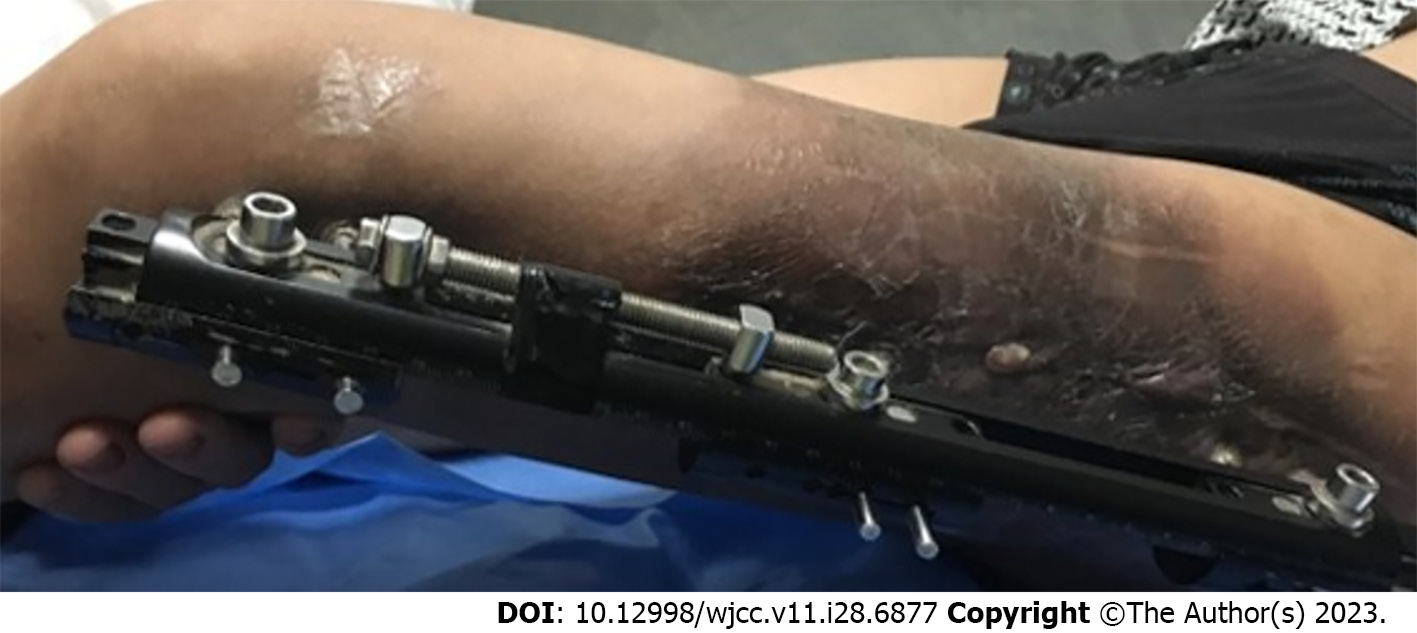
Figure 1 Leathery appearance of the limb after radiotherapy.
Figure 2 X-ray showing nonunion in the middle and upper femur and external fixation.
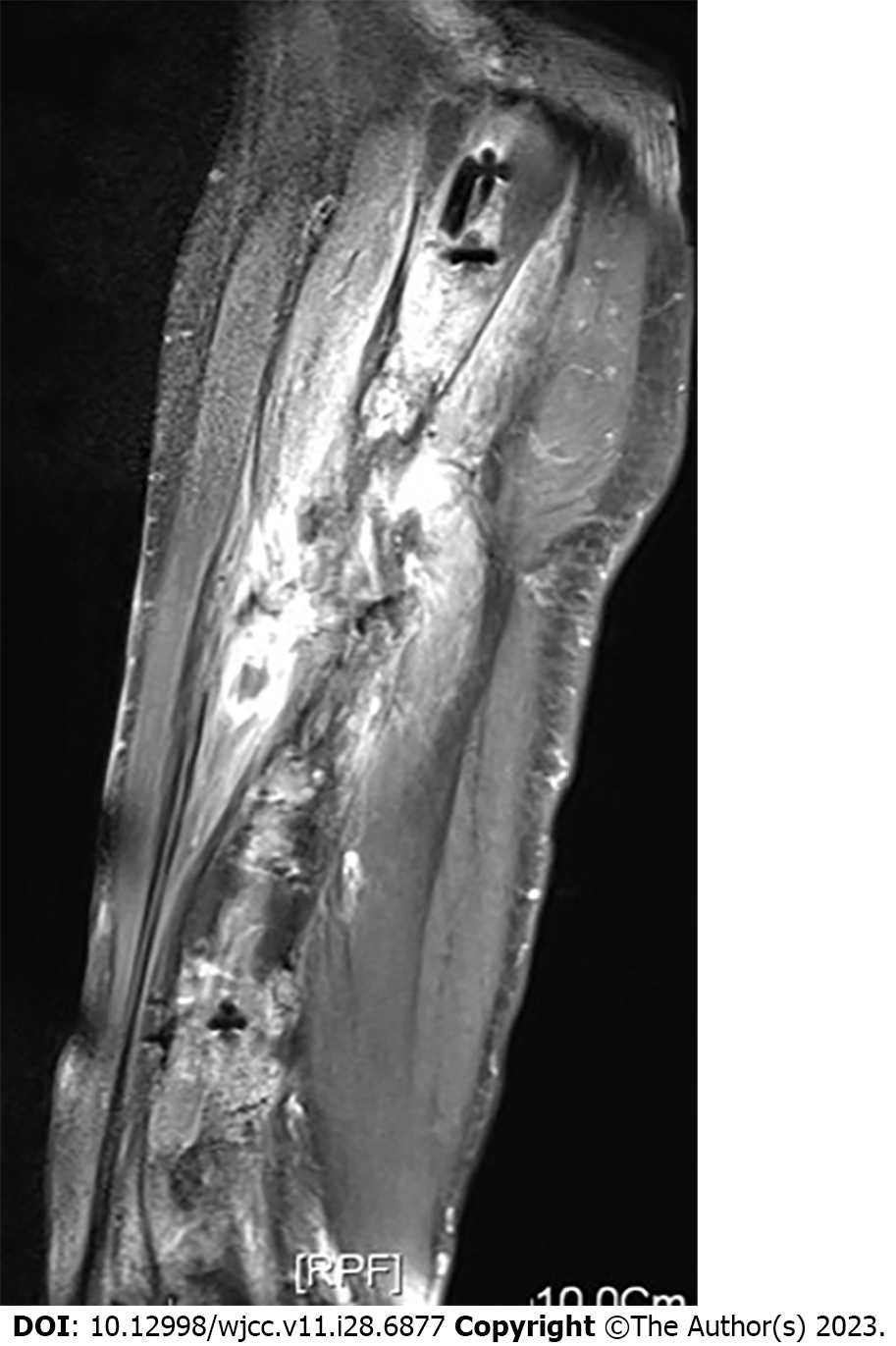
Figure 3 Magnetic resonance imaging showing extensive edema and accumulation of pus at the broken end.
Figure 4 The tumor extensively encircled the femoral vessels and nerves, with extensive involvement in the anterior muscles, minor involvement in the posterior muscles, and no involvement of the sciatic nerve.
Figure 5 The tumor involved the entire femur, up to the greater trochanter.
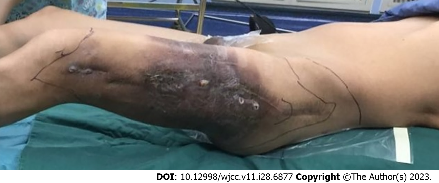
Figure 6 Incisions designed before surgery.
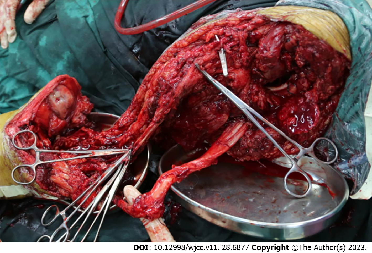
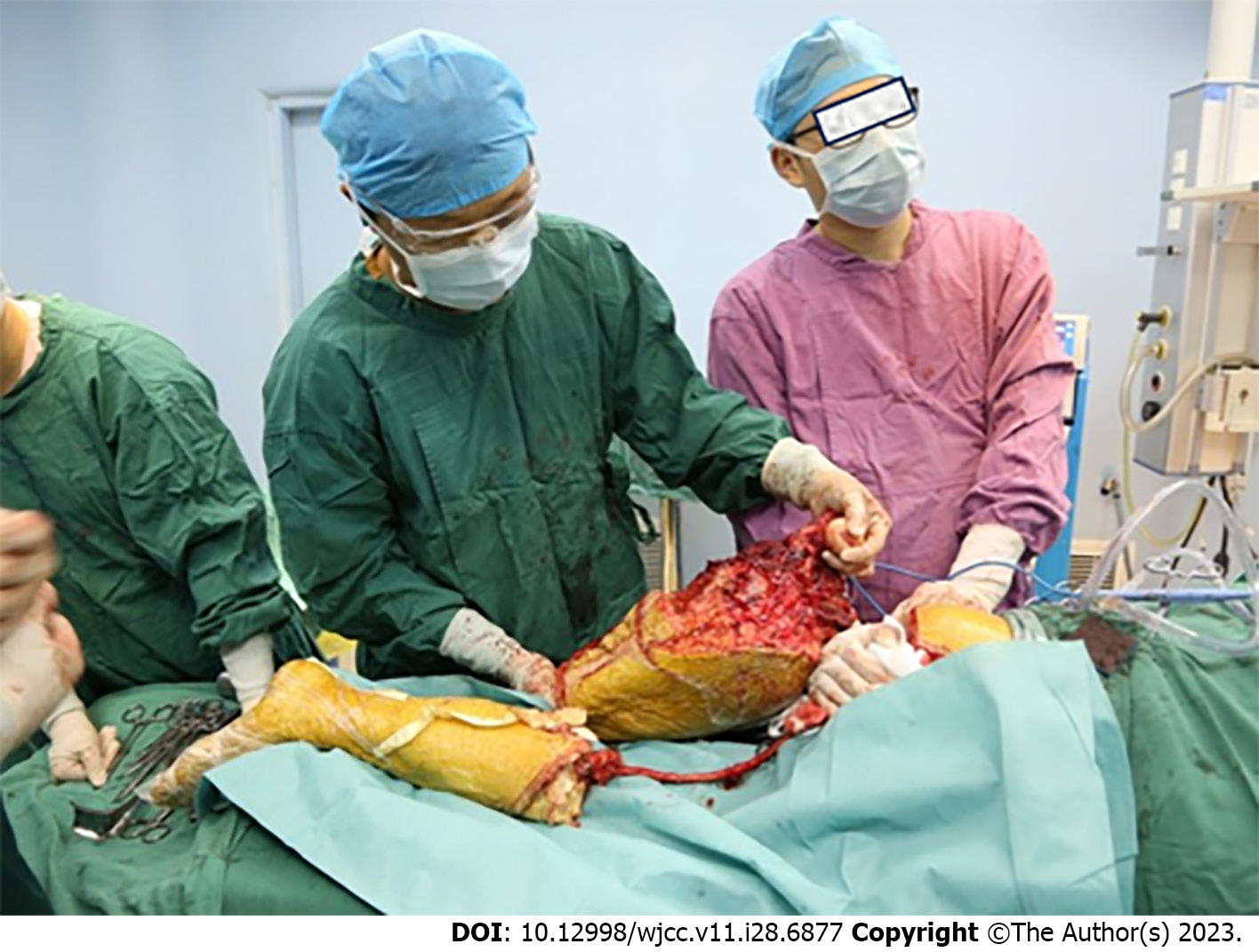
Figure 7 The sciatic nerve was separated.
The procedure created a stenotic tissue flap composed of skin, muscle, sciatic nerve, and femoral vessels.
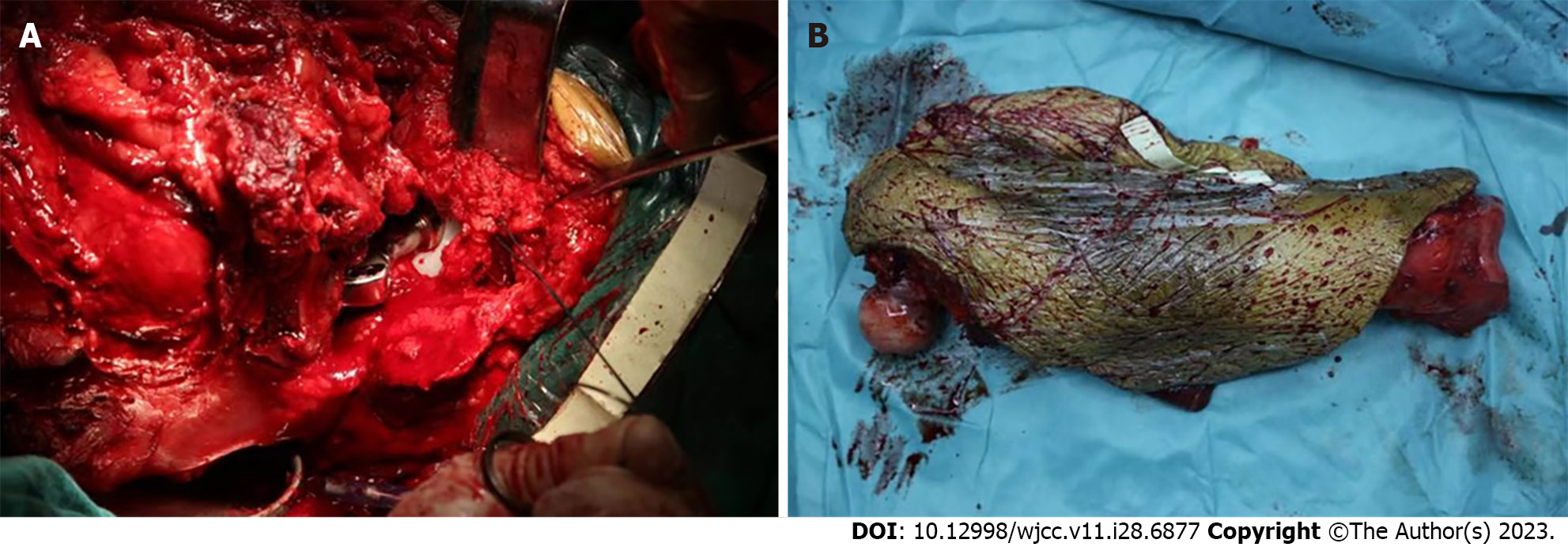
Figure 8 Implants, muscle tissue reconstruction, and amputated femur.
A: Implants and muscle tissue reconstruction; B: Amputated femur.
Figure 9 The whole femur and tumor were removed completely; only the sciatic nerve was preserved.
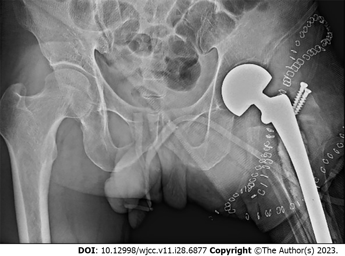
Figure 10 Radiograph of the hip joint indicating that the prosthesis was in a satisfactory position and that the greater trochanter was in a good position with screws.
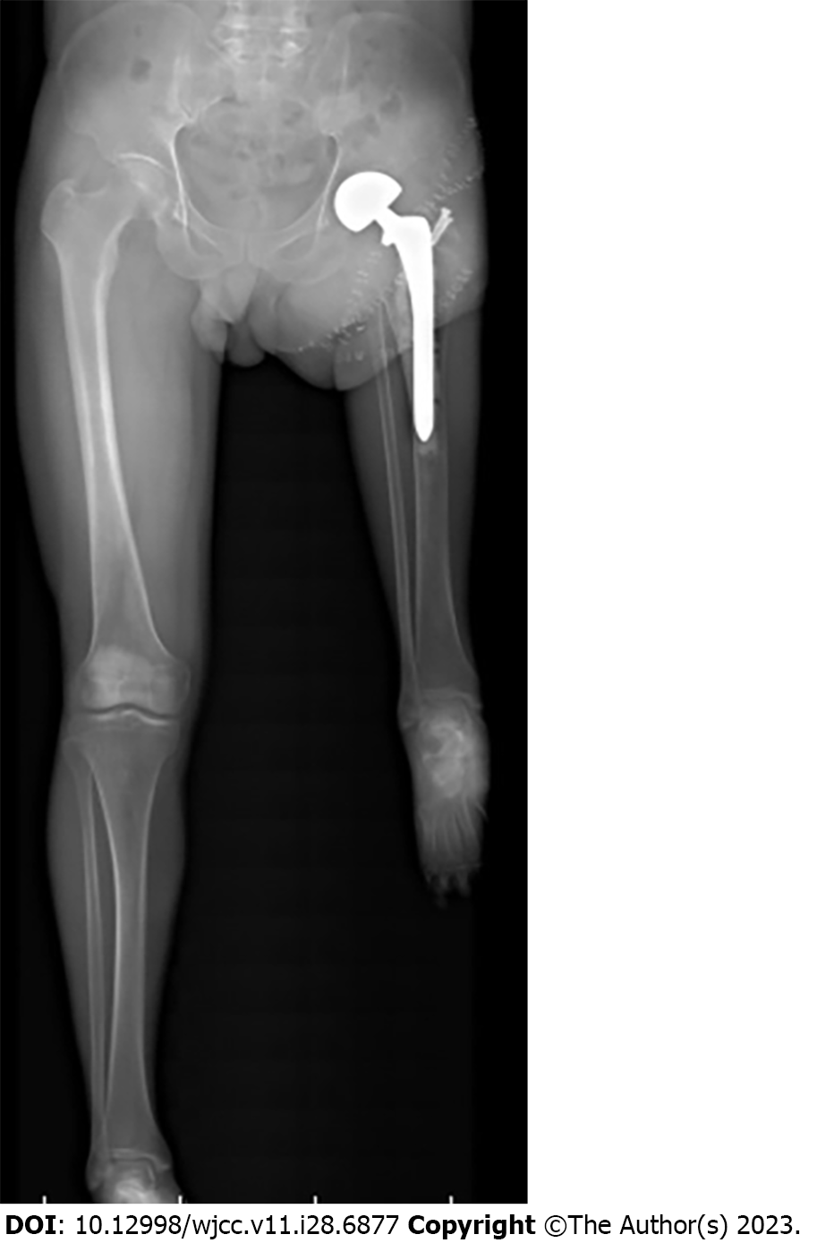
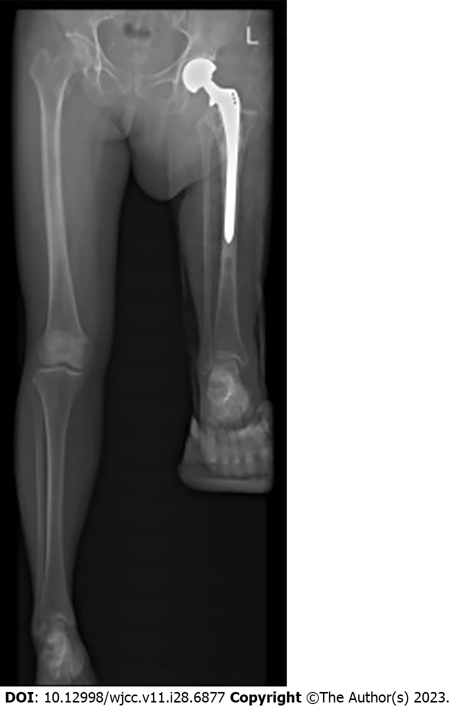
Figure 11 Panoramic image of the lower limbs suggest that the ankle joint of the affected limb is equal to the knee joint of the healthy side.
Figure 12 Full-field radiograph of both lower limbs indicate the satisfactory placement of the prosthesis.
- Citation: Chen ZX, Guo XW, Hong HS, Zhang C, Xie W, Sha M, Ding ZQ. Rotationplasty type BIIIb as an effective alternative to limb salvage procedure in adults: Two case reports. World J Clin Cases 2023; 11(28): 6877-6888
- URL: https://www.wjgnet.com/2307-8960/full/v11/i28/6877.htm
- DOI: https://dx.doi.org/10.12998/wjcc.v11.i28.6877


