Copyright
©The Author(s) 2023.
World J Clin Cases. Oct 6, 2023; 11(28): 6864-6870
Published online Oct 6, 2023. doi: 10.12998/wjcc.v11.i28.6864
Published online Oct 6, 2023. doi: 10.12998/wjcc.v11.i28.6864
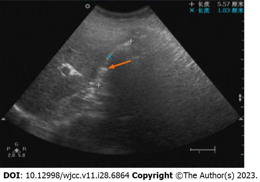
Figure 1 Ultrasound diagnosis before surgery.
Transverse view of the liver showing the gallbladder was filled with a strong hyperechoic mass of uncertain size (arrow).
Figure 2 Ultrasound diagnosis after surgery.
A: Transverse view of the liver showing a smooth liver surface and liver hilum (arrow); B: Transverse view of the gallbladder revealing unclear gallbladder visualization and potential presence of minimal fluid accumulation in the gallbladder area (arrow).
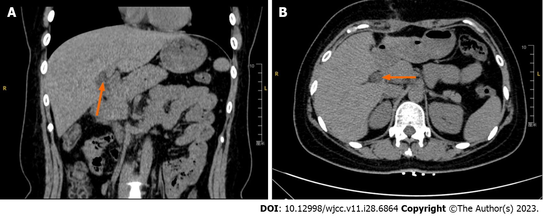
Figure 3 Computed tomography imaging diagnosis.
A: Coronal plane showing dilated intrahepatic bile ducts (arrow); B: Transverse plane demonstrating the absence of a clearly visualized normal gallbladder structure. In the gallbladder fossa region, a linear, slightly higher density shadow was observed (arrow).
Figure 4 Magnetic resonance cholangiopancreatography imaging diagnosis.
A: Coronal view showing indistinct visualization of the linear shadow in the gallbladder fossa (arrow); B: Magnified image at the porta hepatis demonstrating dilation of the bile ducts and irregular thickness of the common bile duct (arrow) with suboptimal local contrast enhancement.
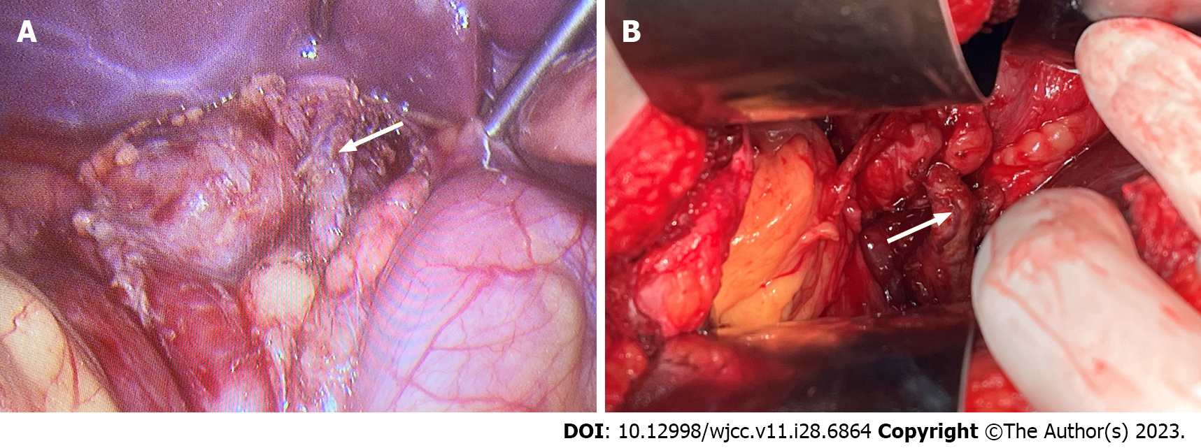
Figure 5 Intraoperative findings and surgical procedure.
A and B: Confirmation of the absence of the gallbladder during surgical exploration. Surgical exploration revealed adhesions between the stomach and liver, which were carefully dissected and separated and dissection of the hepatoduodenal ligament and complete liberation of the common bile duct (arrow).
- Citation: Sun HJ, Ge F, Si Y, Wang Z, Sun HB. Importance of accurate diagnosis of congenital agenesis of the gallbladder from atypical gallbladder stone presentations: A case report. World J Clin Cases 2023; 11(28): 6864-6870
- URL: https://www.wjgnet.com/2307-8960/full/v11/i28/6864.htm
- DOI: https://dx.doi.org/10.12998/wjcc.v11.i28.6864


