Copyright
©The Author(s) 2023.
World J Clin Cases. Oct 6, 2023; 11(28): 6841-6849
Published online Oct 6, 2023. doi: 10.12998/wjcc.v11.i28.6841
Published online Oct 6, 2023. doi: 10.12998/wjcc.v11.i28.6841
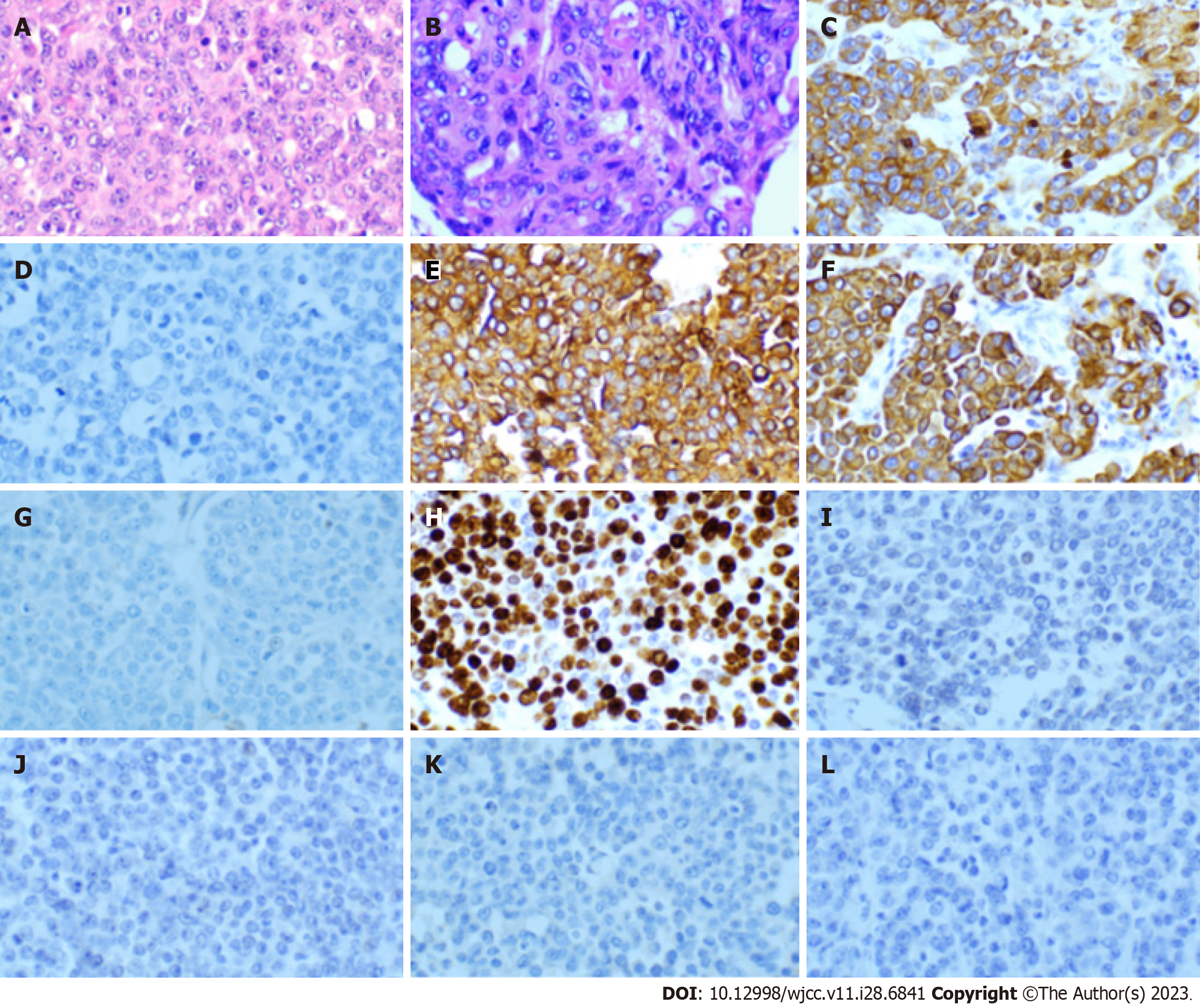
Figure 1 Histological pattern of fine needle puncture of the abdominal subcutaneous nodule.
A and B: Hematoxylin and eosin staining of four sites in the fine needle biopsy specimen showed esophageal squamous cell carcinoma (magnifcation × 40); C-L: Immunohistochemistry for AE1-AE3, CgA (-), CK7 (+), CK34βe (+), GATA3 (-), Ki-67 (index 80%), P63 (-), PAX-8 (-), Syn (-), and TTF-1 (-) in a serial section of the same specimen in shows positive staining predominantly in the tumor tissue (magnification × 40).
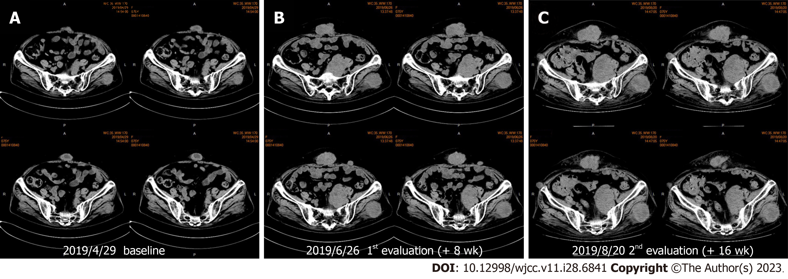
Figure 2 Some subcutaneous nodules and intrapelvic metastases increased significantly, indicating that the patient possibly experienced hyperprogression after immunotherapy.
A: Subcutaneous nodules and intrapelvic nodules two weeks before starting immunotherapy (baseline); B: After 2 cycles of immunological checkpoint inhibitor treatment, abdominal subcutaneous metastases and intrapelvic metastases were significantly increased; C: After 2 wk of pemetrexed chemotherapy, the subcutaneous metastases of the abdominal wall, gluteus maximus metastases, and the intrapelvic metastases did not increase significantly.
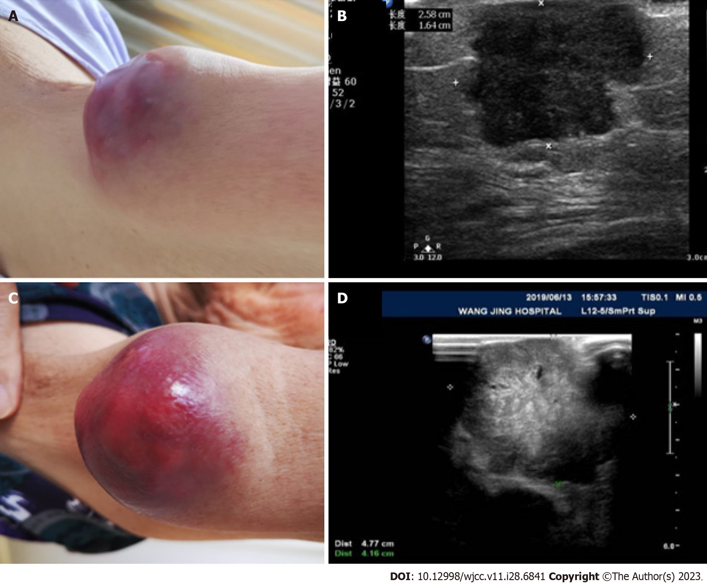
Figure 3 The size of the upper arm metastasis after 2 cycles of immunotherapy increased by 225% compared with the baseline before immunotherapy.
A and B: Baseline: 2.58 mm × 1.64 mm; C and D: 1st evaluation: 4.77 mm × 4.16 mm.
- Citation: Yang HY, Du YX, Hou YJ, Lu DR, Xue P. Hyperprogression after anti-programmed death-1 therapy in a patient with urothelial bladder carcinoma: A case report. World J Clin Cases 2023; 11(28): 6841-6849
- URL: https://www.wjgnet.com/2307-8960/full/v11/i28/6841.htm
- DOI: https://dx.doi.org/10.12998/wjcc.v11.i28.6841


