Copyright
©The Author(s) 2023.
World J Clin Cases. Oct 6, 2023; 11(28): 6792-6796
Published online Oct 6, 2023. doi: 10.12998/wjcc.v11.i28.6792
Published online Oct 6, 2023. doi: 10.12998/wjcc.v11.i28.6792
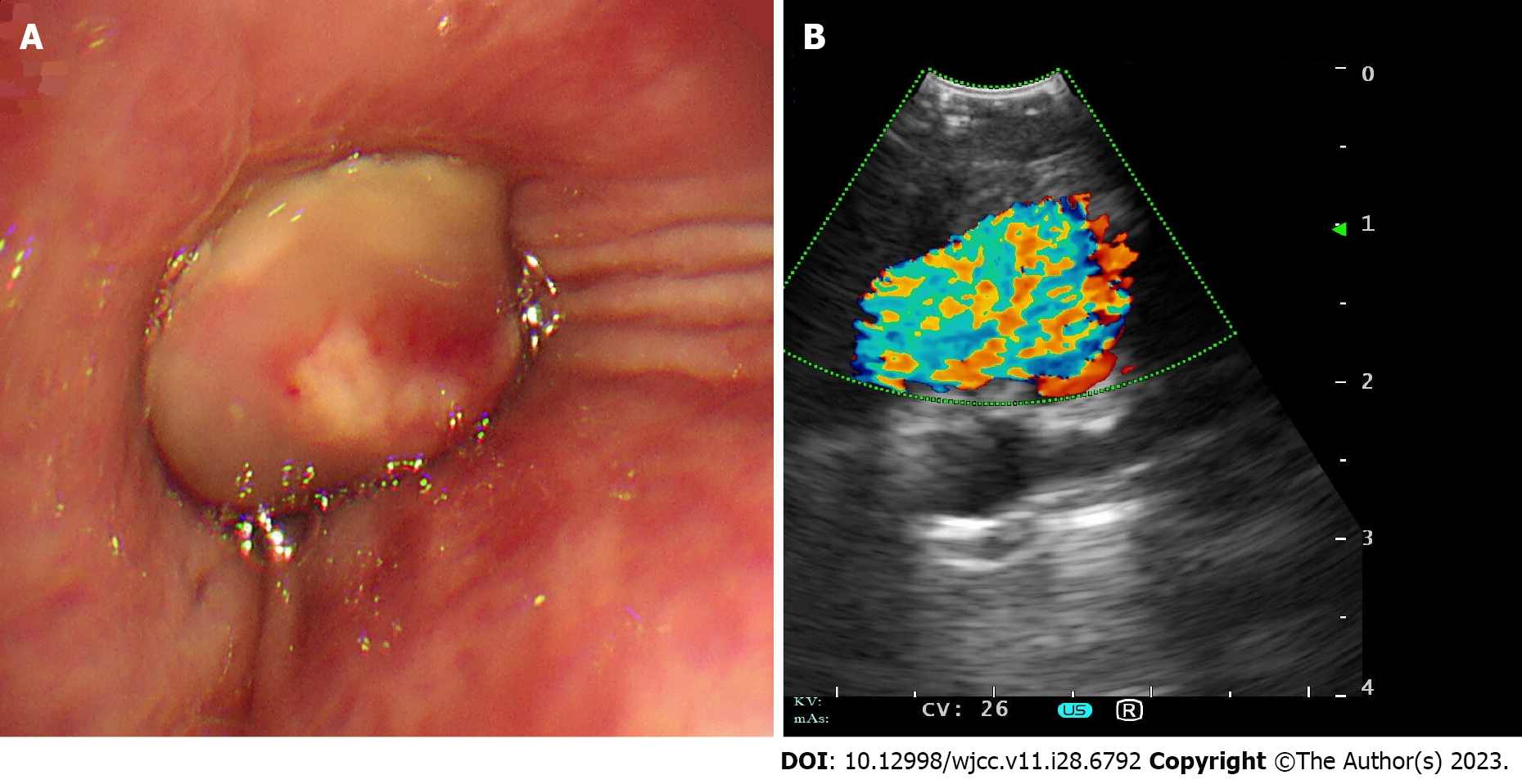
Figure 1 Bronchial examination.
A: Endobronchial lesion visualized under conventional bronchoscopy. It revealed a round, smooth-surfaced mass and flesh colored tumorous protrusion completely blocking the right middle lobe bronchus; B: Endobronchial ultrasonography revealing a vascular lesion.
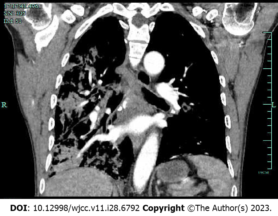
Figure 2 Computed tomography angiography confirming a pulmonary artery aneurysm in the right middle lobe.
- Citation: Li M, Zhu WY, Wu RR, Wang L, Mo MT, Liu SN, Zhu DY, Luo Z. Pulmonary artery aneurysm protruding into the bronchus as an endobronchial mass: A case report. World J Clin Cases 2023; 11(28): 6792-6796
- URL: https://www.wjgnet.com/2307-8960/full/v11/i28/6792.htm
- DOI: https://dx.doi.org/10.12998/wjcc.v11.i28.6792


