Copyright
©The Author(s) 2023.
World J Clin Cases. Oct 6, 2023; 11(28): 6698-6706
Published online Oct 6, 2023. doi: 10.12998/wjcc.v11.i28.6698
Published online Oct 6, 2023. doi: 10.12998/wjcc.v11.i28.6698
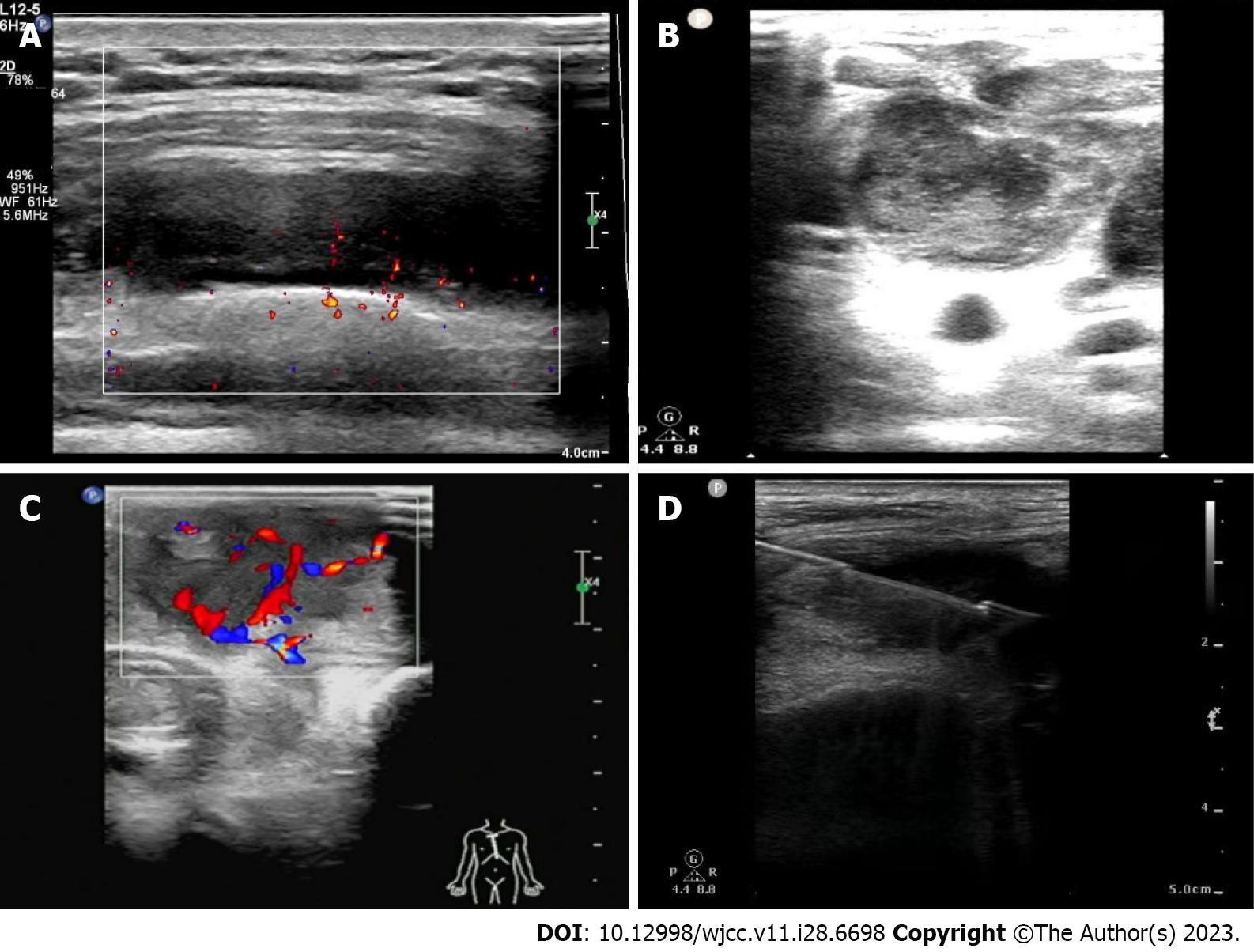
Figure 1 Ultrasound-guided biopsy.
A: Gray scale ultrasound-guided (USG) images clearly reveals the lumps in the chest wall; B: Gray scale USG images clearly reveals the hypoechoic nodules in the chest wall; C: Color Doppler clearly shows the blood flow in a chest wall tuberculosis; D: The ultrasound directs the puncture needle when performing the biopsy.
Figure 2 Receiver operating characteristic curve of pathology and three test methods in the diagnosis of the chest wall tuberculosis.
- Citation: Yan QH, Chi JY, Zhang L, Xue F, Cui J, Kong HL. Value of ultrasound guided biopsy combined with Xpert Mycobacterium tuberculosis/resistance to rifampin assay in the diagnosis of chest wall tuberculosis. World J Clin Cases 2023; 11(28): 6698-6706
- URL: https://www.wjgnet.com/2307-8960/full/v11/i28/6698.htm
- DOI: https://dx.doi.org/10.12998/wjcc.v11.i28.6698


