Copyright
©The Author(s) 2023.
World J Clin Cases. Sep 26, 2023; 11(27): 6646-6652
Published online Sep 26, 2023. doi: 10.12998/wjcc.v11.i27.6646
Published online Sep 26, 2023. doi: 10.12998/wjcc.v11.i27.6646
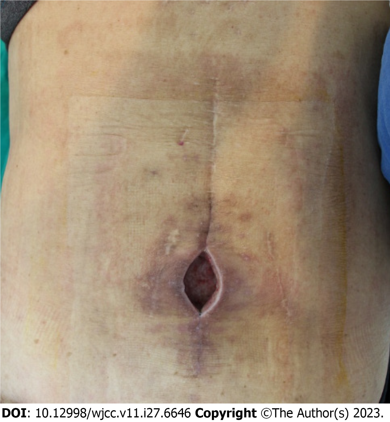
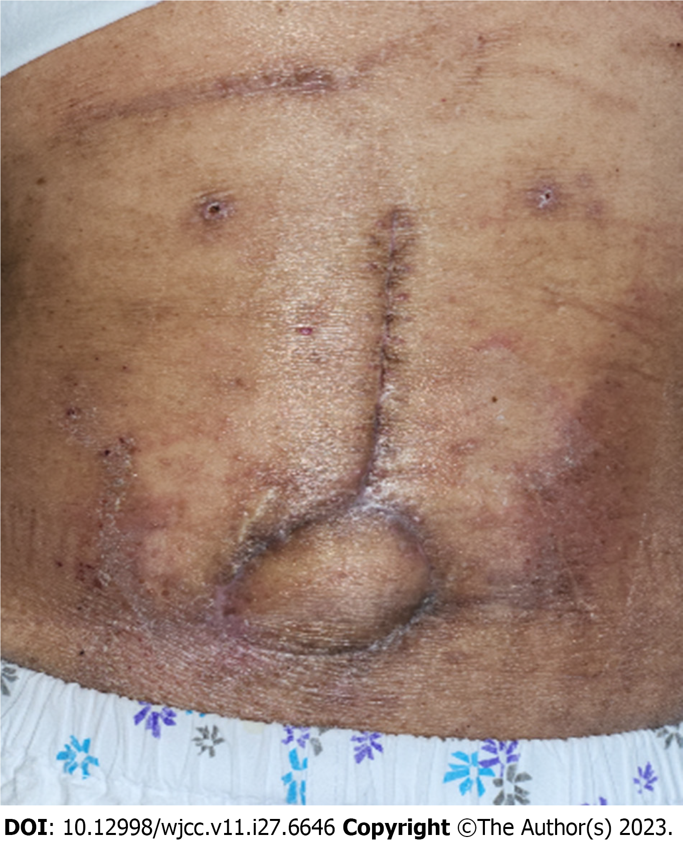
Figure 1 A 75-year-old man with a chronic wound on the lower back.
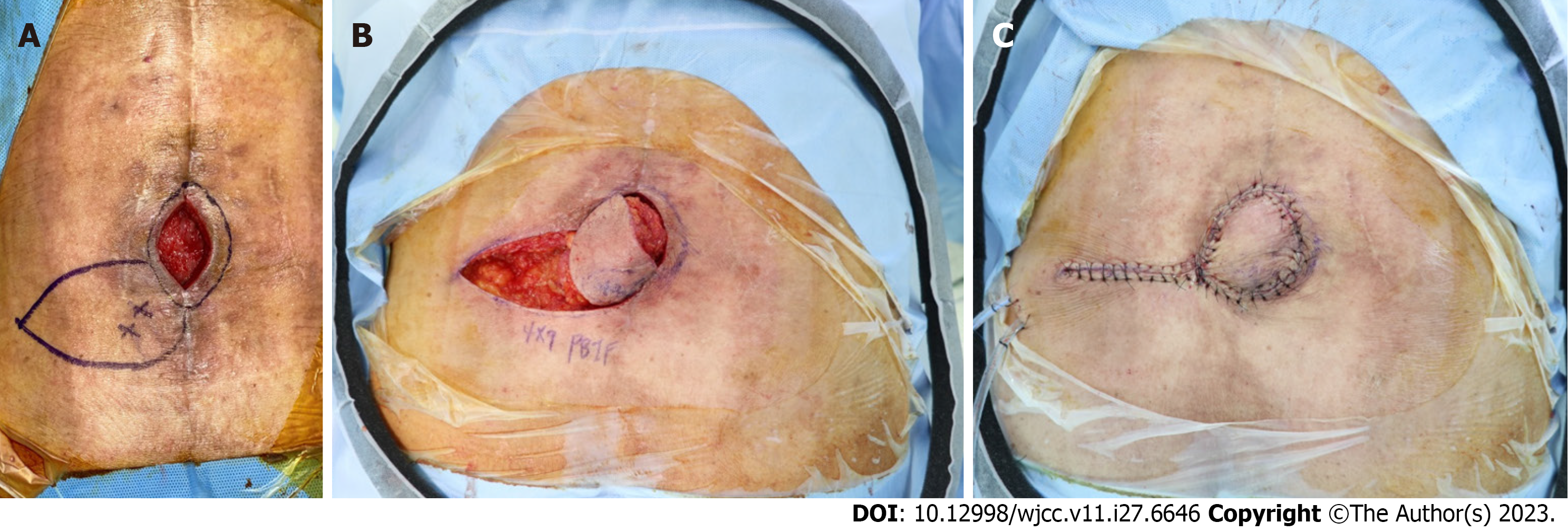
Figure 2 Intraoperative photographs.
A: A 4 cm × 9 cm sized perforator-based island flap was designed; B: Island-type insetting was tried after defatting; C: Authors confirmed no tension to the perforator before skin closure.
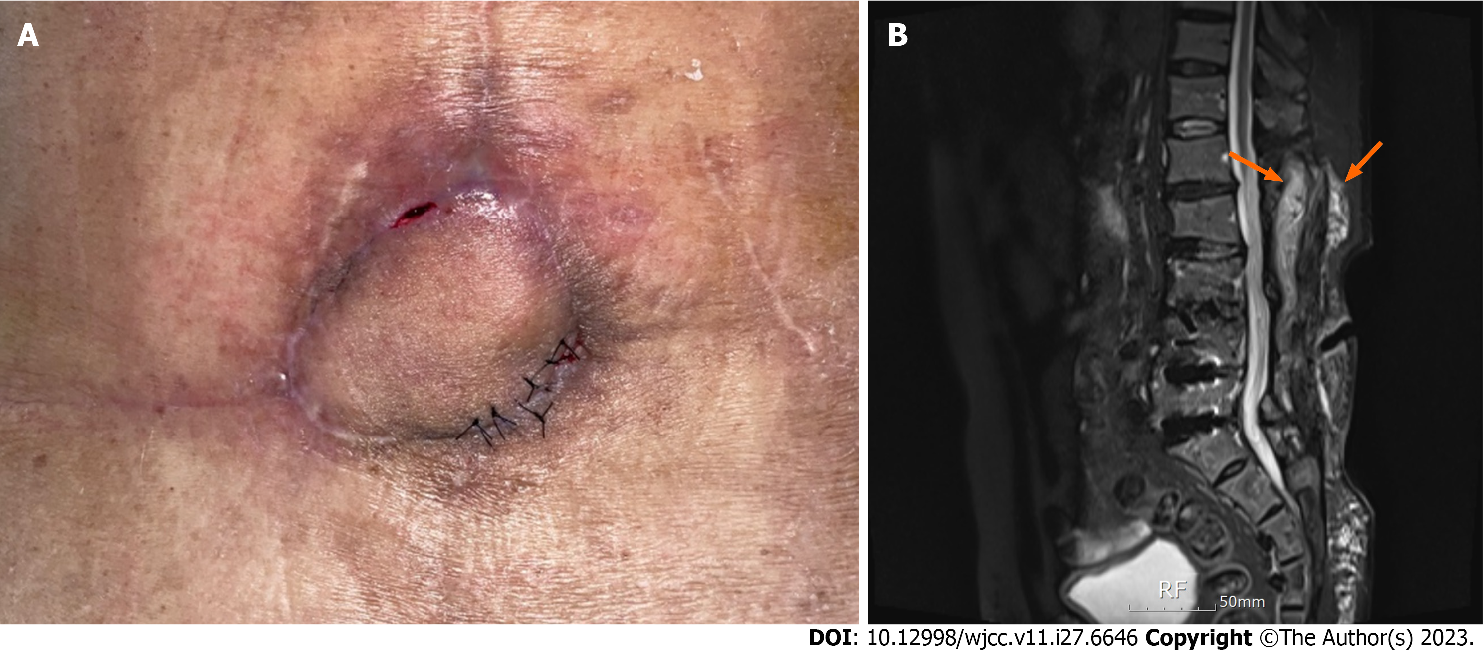
Figure 3 Delayed infection was noticed.
A: Another abscess formation was noticed in the 11 o’clock region of the flap; B: 1.5T enhanced L-spine magnetic resonance imaging T2 imaging study revealed suspicious soft tissue phlegmon (orange arrow) along the interspinous reconstruction wire.
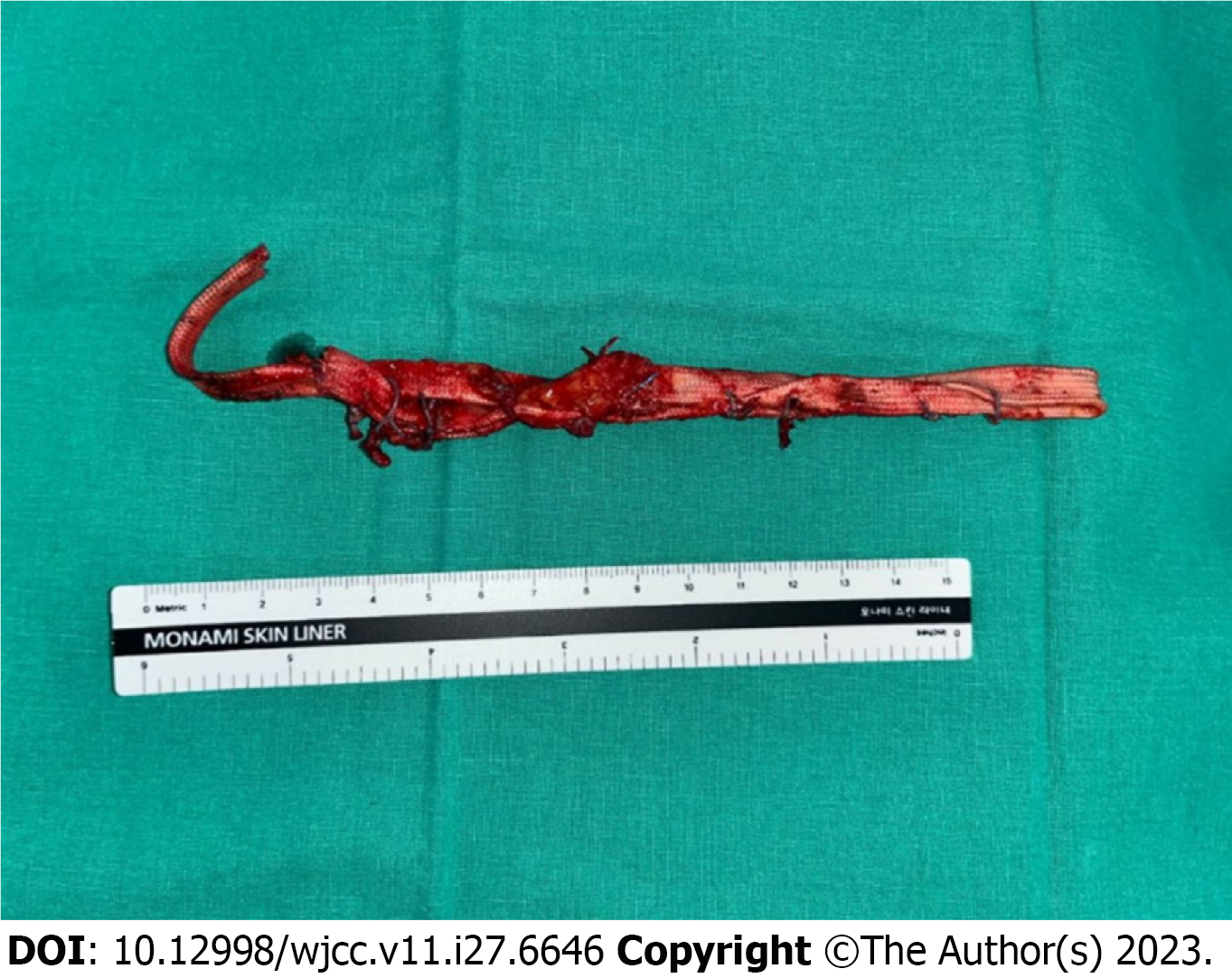
Figure 4 Infected interspinous reconstruction wire was removed by the spine surgeon.
Figure 5 The patient was discharged without further complications.
- Citation: Kim D, Lim S, Eo S, Yoon JS. Reconstruction of the lower back wound with delayed infection after spinal surgery: A case report. World J Clin Cases 2023; 11(27): 6646-6652
- URL: https://www.wjgnet.com/2307-8960/full/v11/i27/6646.htm
- DOI: https://dx.doi.org/10.12998/wjcc.v11.i27.6646


