Copyright
©The Author(s) 2023.
World J Clin Cases. Sep 26, 2023; 11(27): 6613-6617
Published online Sep 26, 2023. doi: 10.12998/wjcc.v11.i27.6613
Published online Sep 26, 2023. doi: 10.12998/wjcc.v11.i27.6613
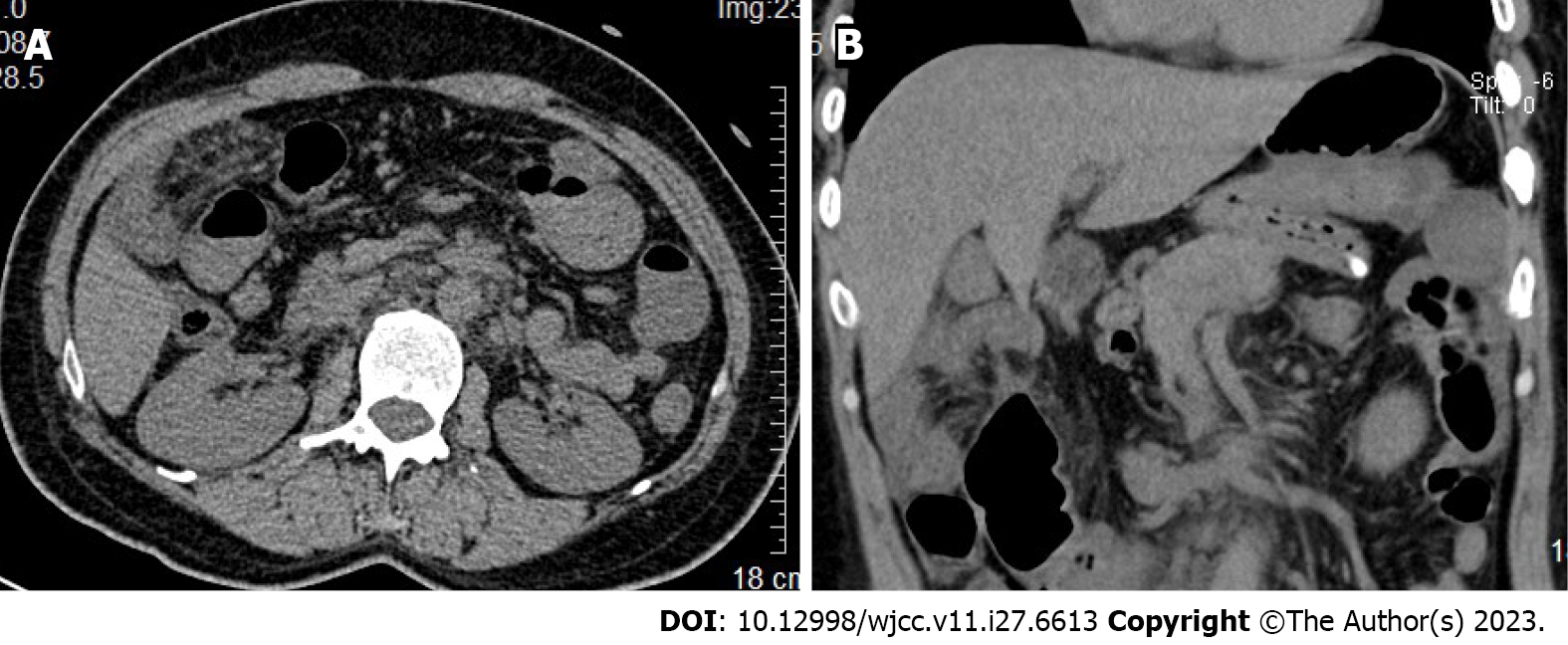
Figure 1 Abdominal computed tomography findings.
A: Transverse view; B: Sagittal view. The small intestine was locally dilated in the abdominal cavity and the intestinal tube wall was thickened. Above the transverse colon, the small intestine and mesangium were partially thickened, and edema of the tube wall was found around the gallbladder.
Figure 2 Intraoperative findings.
A: The yellow dotted line shows herniation into the incarcerated small intestine. The black arrow shows the hepatoduodenal adhesions; B: The blue dotted line shows the transverse mesocolic defect.
Figure 3
Timeline.
- Citation: Zhang C, Guo DF, Lin F, Zhan WF, Lin JY, Lv GF. Transverse mesocolic hernia with intestinal obstruction as a rare cause of acute abdomen in adults: A case report. World J Clin Cases 2023; 11(27): 6613-6617
- URL: https://www.wjgnet.com/2307-8960/full/v11/i27/6613.htm
- DOI: https://dx.doi.org/10.12998/wjcc.v11.i27.6613


