Copyright
©The Author(s) 2023.
World J Clin Cases. Sep 26, 2023; 11(27): 6592-6596
Published online Sep 26, 2023. doi: 10.12998/wjcc.v11.i27.6592
Published online Sep 26, 2023. doi: 10.12998/wjcc.v11.i27.6592
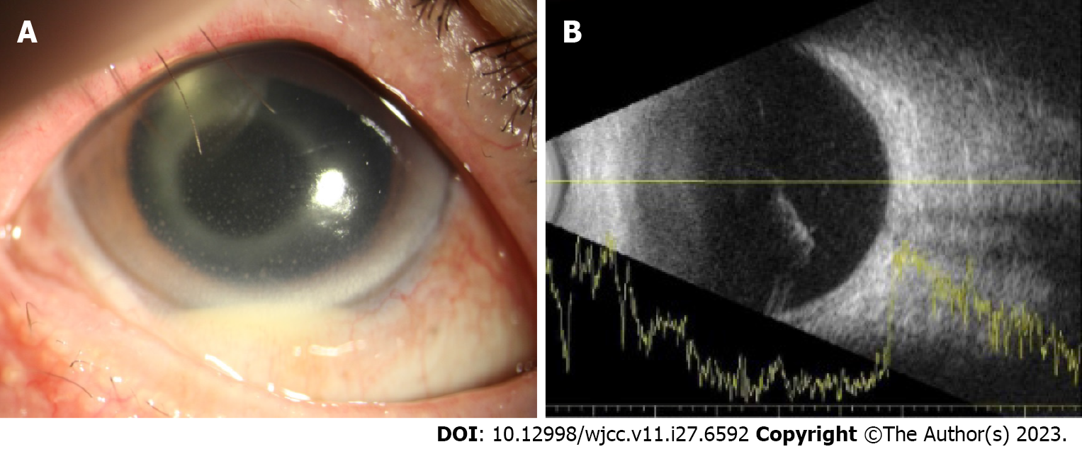
Figure 1 Anterior segment photograph and ultrasonogram at initial presentation in a patient with a history of cataract surgery 4 mo prior.
A: Conjunctival injection, keratic precipitates, and hypopyon formation in the anterior chamber; B: Heterogeneous vitreous opacity is evident in the B-scan ultrasonogram.
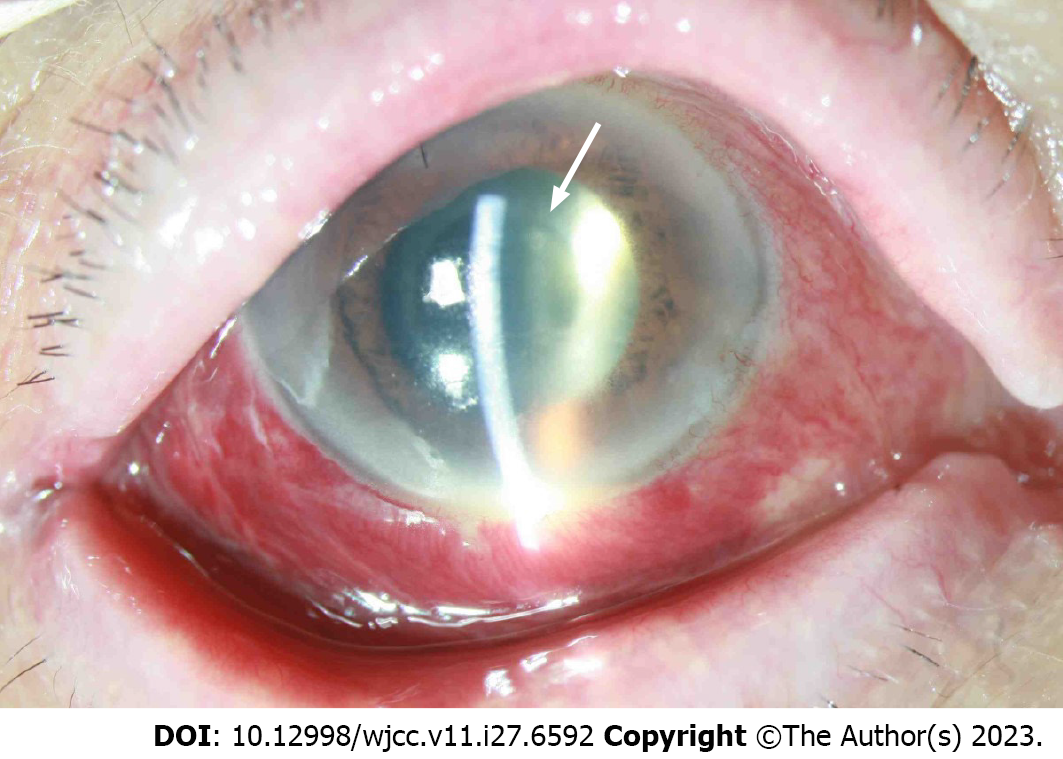
Figure 2 Anterior segment photograph on the day of the second operation.
Whitish plaque was observed in the lens capsule (white arrow). Anterior chamber cell grade increased to 4+ and hypopyon reappeared.
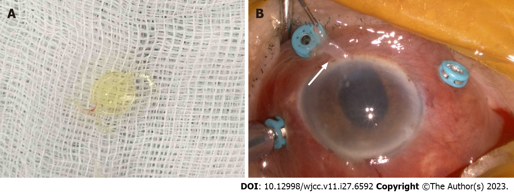
Figure 3 The intraocular lens and capsule (white arrow) were removed during the second operation.
Micrococcus luteus was cultured. A: Intraocular lens; B: Capsule.
- Citation: Nam KY, Lee HW. Delayed-onset micrococcus luteus-induced postoperative endophthalmitis several months after cataract surgery: A case report. World J Clin Cases 2023; 11(27): 6592-6596
- URL: https://www.wjgnet.com/2307-8960/full/v11/i27/6592.htm
- DOI: https://dx.doi.org/10.12998/wjcc.v11.i27.6592


