Copyright
©The Author(s) 2023.
World J Clin Cases. Sep 26, 2023; 11(27): 6587-6591
Published online Sep 26, 2023. doi: 10.12998/wjcc.v11.i27.6587
Published online Sep 26, 2023. doi: 10.12998/wjcc.v11.i27.6587
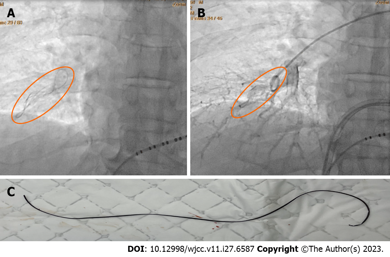
Figure 1 Foreign body.
A: A foreign body appeared in the right lower lung field; B: Pulmonary angiography showed a foreign body inside the right lower pulmonary artery; C: The foreign body was a line-like substance about 15 cm long.
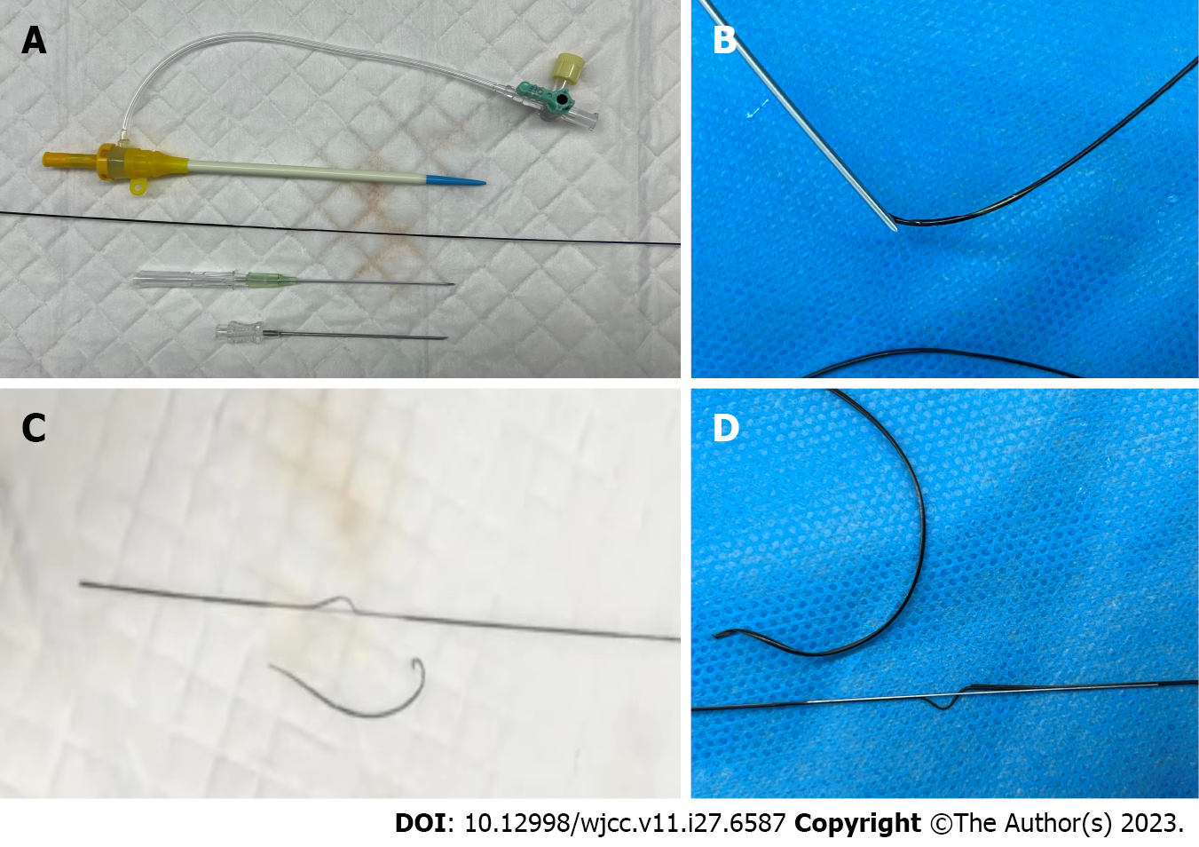
Figure 2 Instruments for femoral vein puncture.
A: Needle sheath system (11F, Terumo, Japan); B-D: The formation of the foreign body.
- Citation: Yan R, Lei XY, Li J, Jia LL, Wang HX. Removal of a pulmonary artery foreign body during pulse ablation in a patient with atrial fibrillation: A case report. World J Clin Cases 2023; 11(27): 6587-6591
- URL: https://www.wjgnet.com/2307-8960/full/v11/i27/6587.htm
- DOI: https://dx.doi.org/10.12998/wjcc.v11.i27.6587


