Copyright
©The Author(s) 2023.
World J Clin Cases. Sep 26, 2023; 11(27): 6579-6586
Published online Sep 26, 2023. doi: 10.12998/wjcc.v11.i27.6579
Published online Sep 26, 2023. doi: 10.12998/wjcc.v11.i27.6579
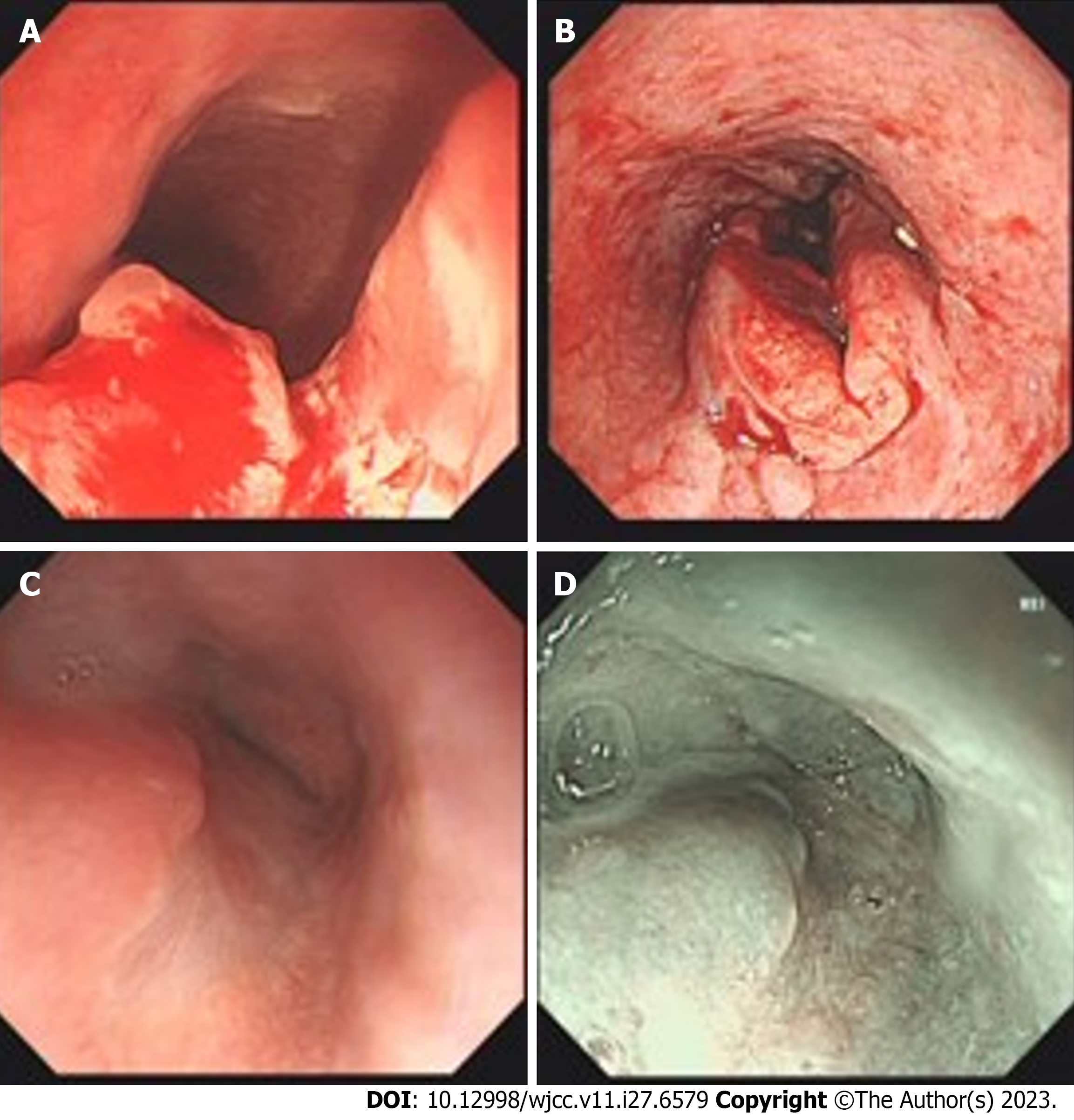
Figure 1 Results of endoscopy.
A: The lesion located at the middle and lower parts of the esophagus before therapy; B: Image after endoscopic biopsy; C: Tumor shrank or disappeared after 3 mo of combined toripalimab and anlotinib therapy; D: Staining magnifying endoscope after 3 mo of the combination therapy.
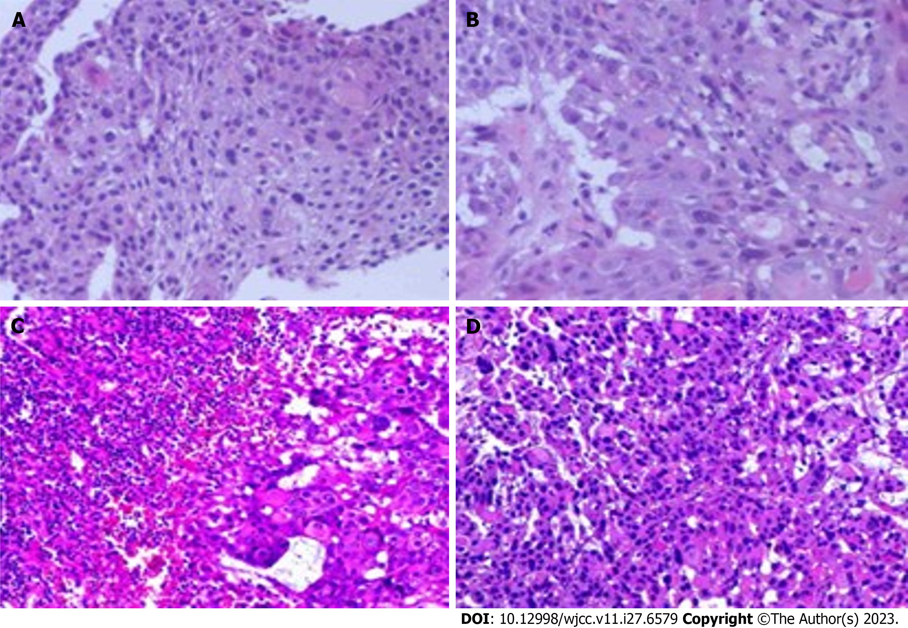
Figure 2 Histopathological examination by hematoxylin-eosin staining.
A and B: Heterogeneous proliferation of tissue squamous epithelium. The heterogeneous cells break through the basement membrane and infiltrate below the mesenchyme (40 ×, 200 ×); C and D: The squamous epithelial cells were markedly heterogeneous and showed infiltrative growth, and a little lymphoid tissue was seen in their periphery (40 ×, 200 ×).
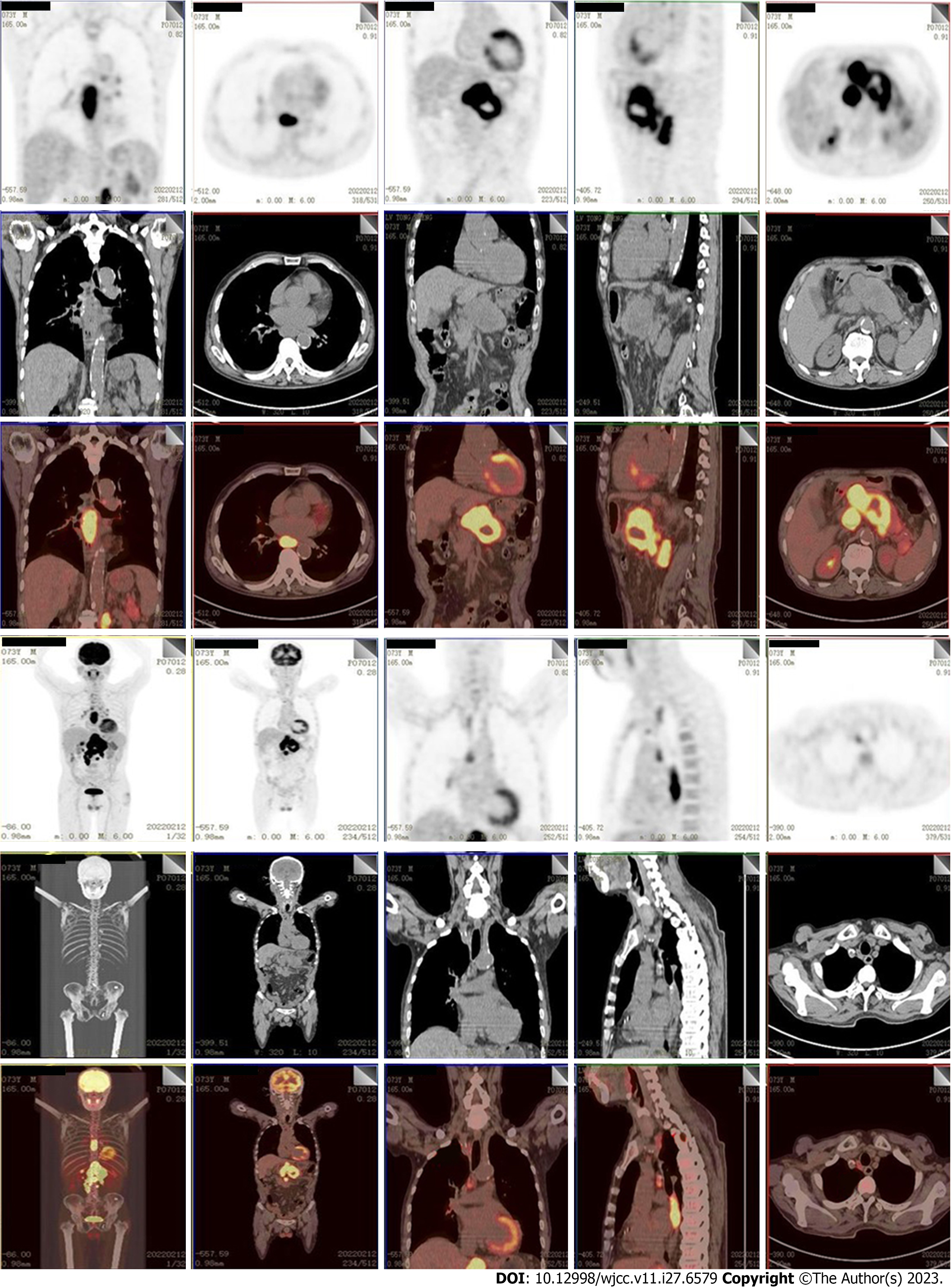
Figure 3 Positron emission tomography/computed tomography scan images before the treatment.
Two hypermetabolic shadows were respectively seen in each of the lower and middle esophagus (T6-9 level) and the body of the pancreas, which were diagnosed as malignancies. Multiple enlarged and hypermetabolic lymph nodes were located in the right mediastinum, hilum, surrounding pancreas, retroperitoneum, and the surrounding hepatogastric ligament.
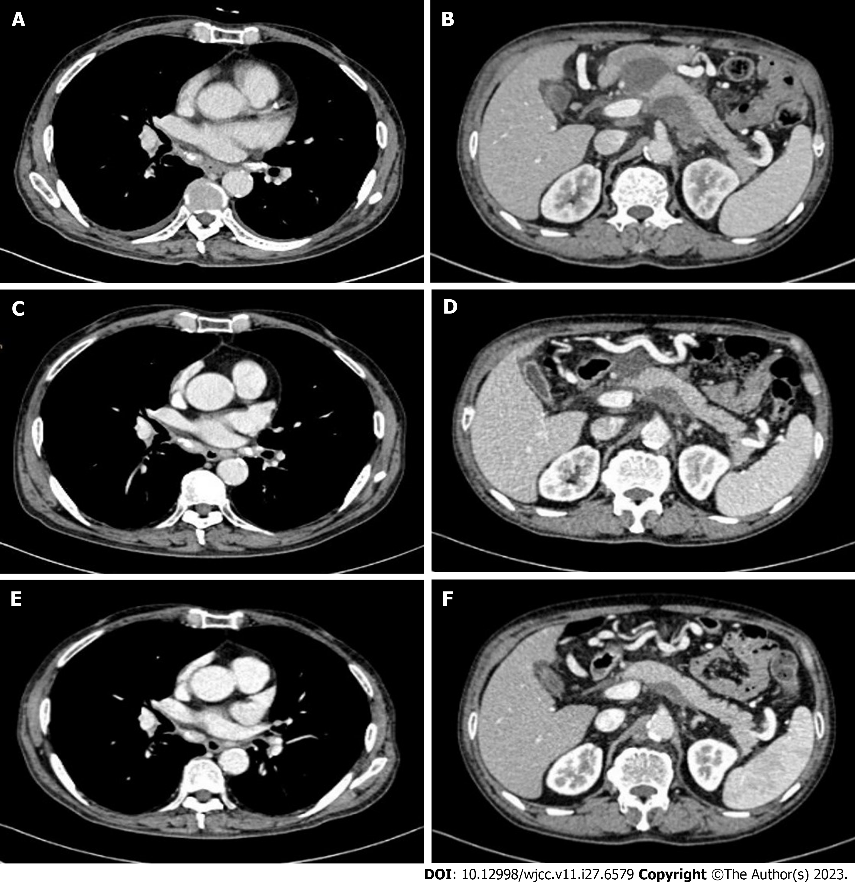
Figure 4 Enhanced computed tomography scan images during therapy.
A and B: 1 mo after combined toripalimab and anlotinib therapy, the esophagus lesion and metastatic lymph nodes shrunk; C and D: The tumor and metastatic lymph nodes were disappeared after 3 mo of combination therapy; E and F: The tumor and metastatic lymph nodes have been disappeared for up to 14 mo since the initiation of combination therapy.
- Citation: Chen SC, Ma DH, Zhong JJ. Combination therapy with toripalimab and anlotinib in advanced esophageal squamous cell carcinoma: A case report. World J Clin Cases 2023; 11(27): 6579-6586
- URL: https://www.wjgnet.com/2307-8960/full/v11/i27/6579.htm
- DOI: https://dx.doi.org/10.12998/wjcc.v11.i27.6579


