Copyright
©The Author(s) 2023.
World J Clin Cases. Sep 26, 2023; 11(27): 6565-6572
Published online Sep 26, 2023. doi: 10.12998/wjcc.v11.i27.6565
Published online Sep 26, 2023. doi: 10.12998/wjcc.v11.i27.6565
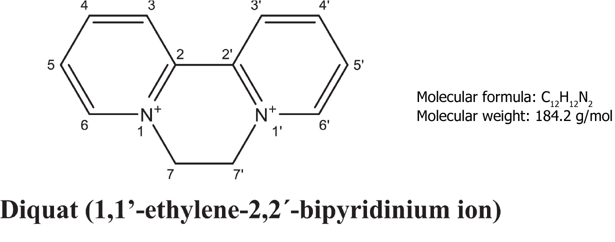
Figure 1 The molecular structure and molecular weight of diquat.
Citation: Magalhães N, Carvalho F, Dinis-Oliveira RJ. Human and experimental toxicology of diquat poisoning: Toxicokinetics, mechanisms of toxicity, clinical features, and treatment. Hum Exp Toxicol 2018; 37: 1131-1160[1]. Published by SAGE Publications. The authors have obtained the permission for figure using from the SAGE Publications. Copyright© The Authors 2018. (Supplementary material).
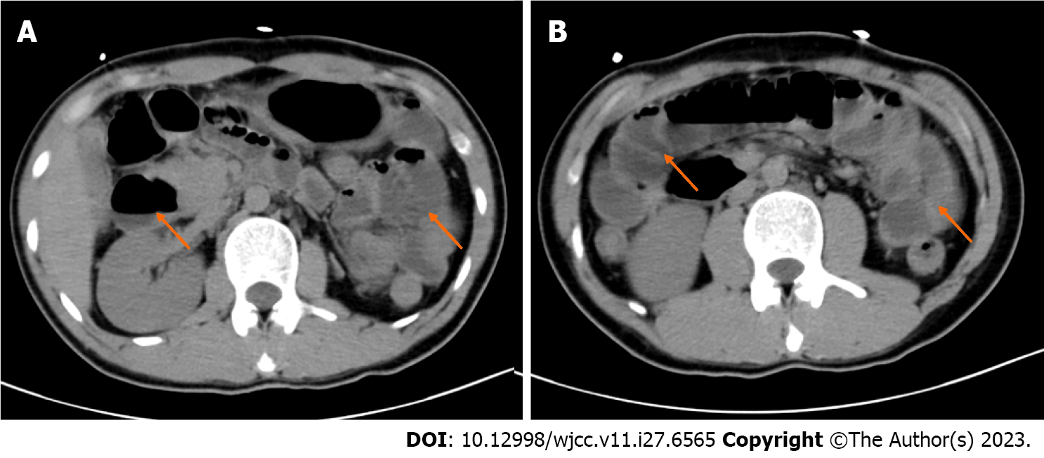
Figure 2 Imaging examinations.
A and B: Abdominal computed tomography of the patient.
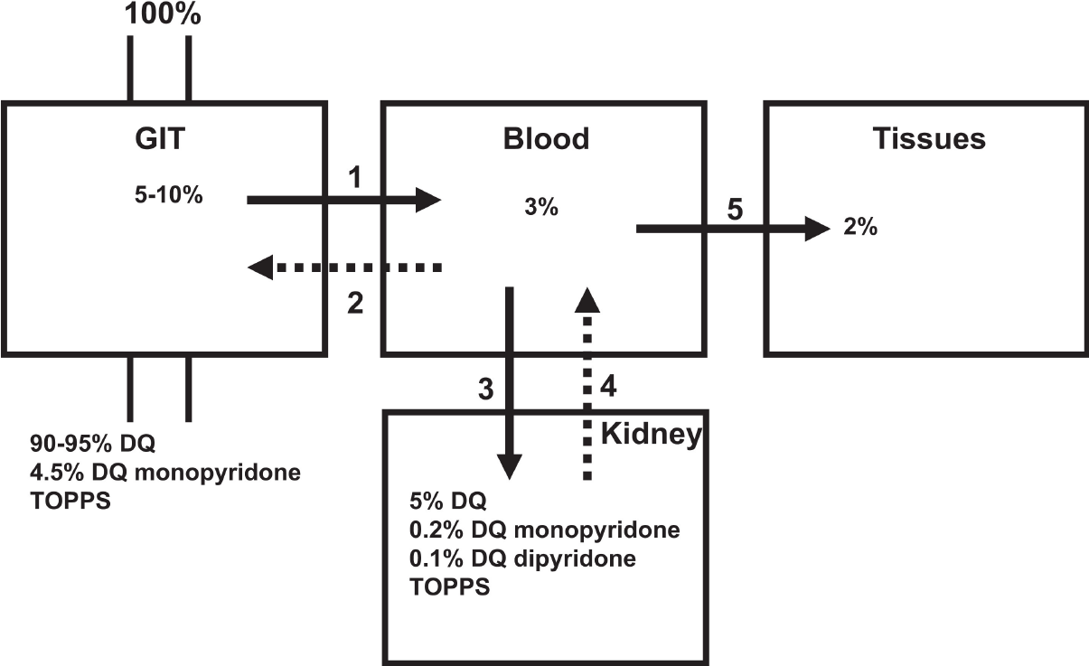
Figure 3 Model course of toxicokinetics of oral diquat ingestion.
Citation: Magalhães N, Carvalho F, Dinis-Oliveira RJ. Human and experimental toxicology of diquat poisoning: Toxicokinetics, mechanisms of toxicity, clinical features, and treatment. Hum Exp Toxicol 2018; 37: 1131-1160[1]. Published by SAGE Publications. The authors have obtained the permission for figure using from the SAGE Publications. Copyright© The Authors 2018. (Supplementary material).
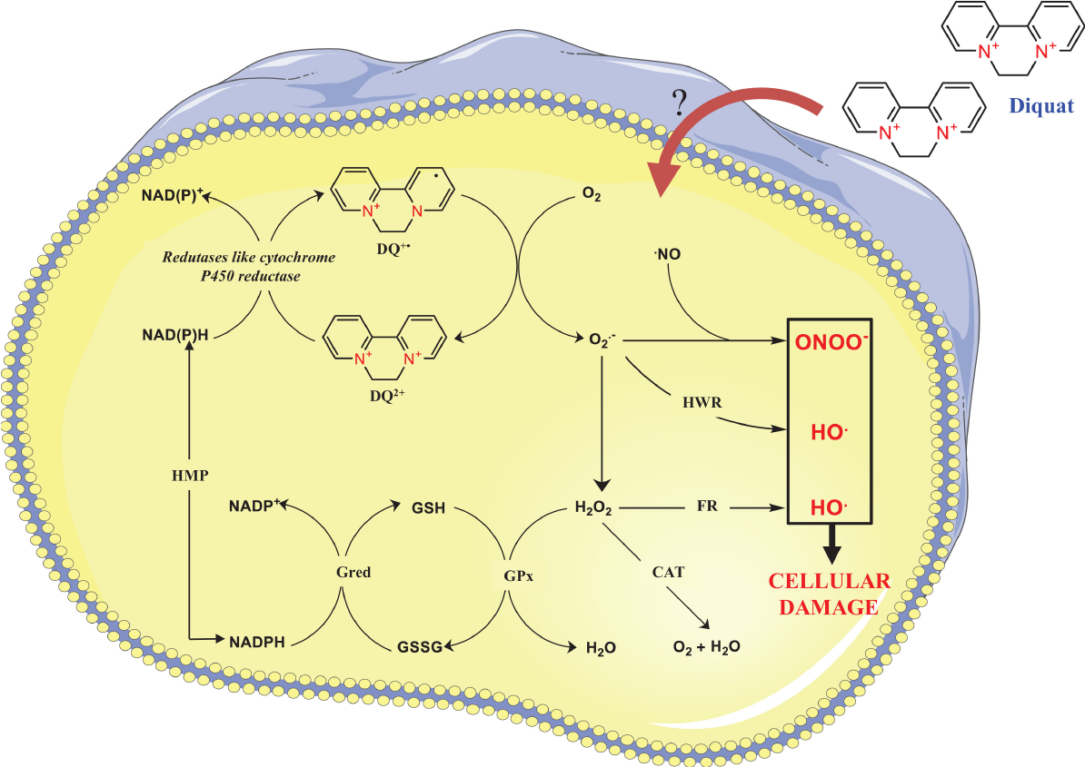
Figure 4 Schematic representation of the redox cycling of diquat and reactive oxygen species.
Citation: Magalhães N, Carvalho F, Dinis-Oliveira RJ. Human and experimental toxicology of diquat poisoning: Toxicokinetics, mechanisms of toxicity, clinical features, and treatment. Hum Exp Toxicol 2018; 37: 1131-1160[1]. Published by SAGE Publications. The authors have obtained the permission for figure using from the SAGE Publications. Copyright© The Authors 2018. (Supplementary material).
- Citation: Fan CY, Zhang CG, Zhang PS, Chen Y, He JQ, Yin H, Gong XJ. Acute diquat poisoning case with multiorgan failure and a literature review: A case report. World J Clin Cases 2023; 11(27): 6565-6572
- URL: https://www.wjgnet.com/2307-8960/full/v11/i27/6565.htm
- DOI: https://dx.doi.org/10.12998/wjcc.v11.i27.6565


