Copyright
©The Author(s) 2023.
World J Clin Cases. Sep 26, 2023; 11(27): 6543-6550
Published online Sep 26, 2023. doi: 10.12998/wjcc.v11.i27.6543
Published online Sep 26, 2023. doi: 10.12998/wjcc.v11.i27.6543
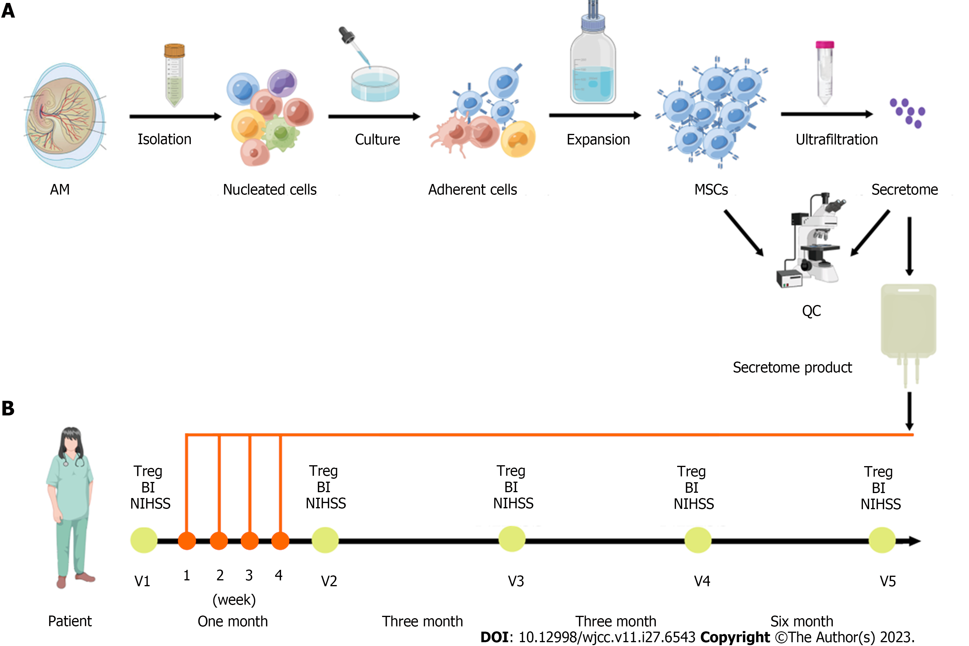
Figure 1 Schematic diagram on clinical-grade manufacturing process of the mesenchymal stromal cell-secretome product and schedule of the treatments of the mesenchymal stromal cell-secretome.
A: Amniotic membrane (AM) mesenchymal stromal cells (MSCs) were isolated, cultured, and expanded, and quality control (QC) evaluated cell characterization, surface markers, differentiation potential, presence of fungi and bacteria, and endotoxin level. The conditioned medium was concentrated by centrifugation using ultrafiltration units with a 3-kDa cutoff. The concentrated media was analyzed for protein concentration and for secreted cytokines content; B: Schematic representation of the follow-up visits (V) before (V1) and after the treatments of the MSC-secretome (V2-V5). The patient was enrolled in March 2021 and received four infusions of 50 mg MSC-secretome once a week for 4 wk. The clinical and laboratory evaluation of the patient was checked at each follow-up visit. BI: Barthel index; NIHSS: National Institute of Health Stroke Scale; Treg: Regulatory T cells.
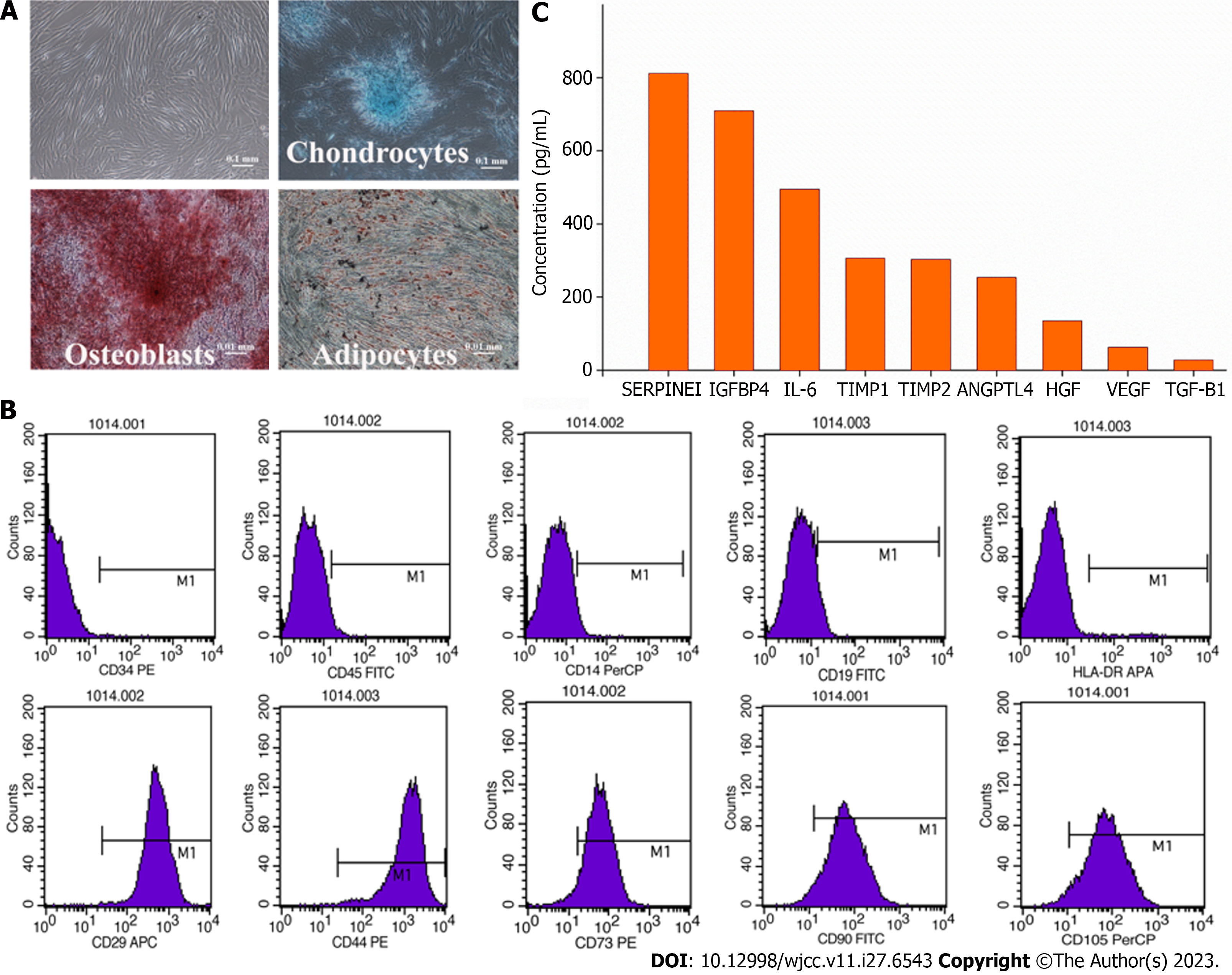
Figure 2 Quality control of amniotic membrane mesenchymal stromal cells and the mesenchymal stromal cell-secretome.
A: Cell characterization and differentiation potential. Amniotic membrane-mesenchymal stromal cells were characterized by their fibroblast-like morphology and their differentiation potential towards osteoblasts, chondrocytes, and adipocytes in special induction medium; B: Surface markers. The mniotic membrane-mesenchymal stromal cells showed a high expression of CD29, CD44, CD73, CD90, CD105 and low expression of CD14, CD19, CD34, CD45, and HLA-DR markers; C: Cytokine concentration. ANGPTL4: Angiopoietin-like 4; HGF: Hepatocyte growth factor; IGFBP4: Insulin-like growth factor binding protein 4; IL-6: Interleukin-6; TIMP1: Tissue inhibitors of metalloproteinase 1; SERPINE1: Serine protease inhibitor clade E member 1; TGF-β1: Transforming growth factor-beta 1; TIMP-2: Tissue inhibitors of metalloproteinase 2; VEGF: Vascular endothelial growth factor.
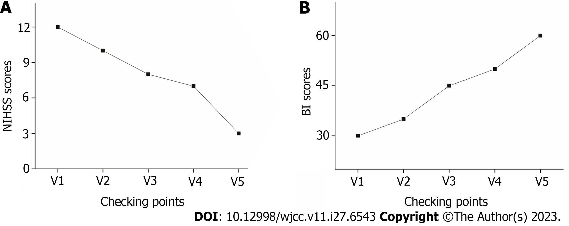
Figure 3 Neurological function before and after mesenchymal stromal cell-secretome transplantation.
A: National Institutes of Health Stroke Scale (NIHSS) score; B: Barthel index (BI) score.
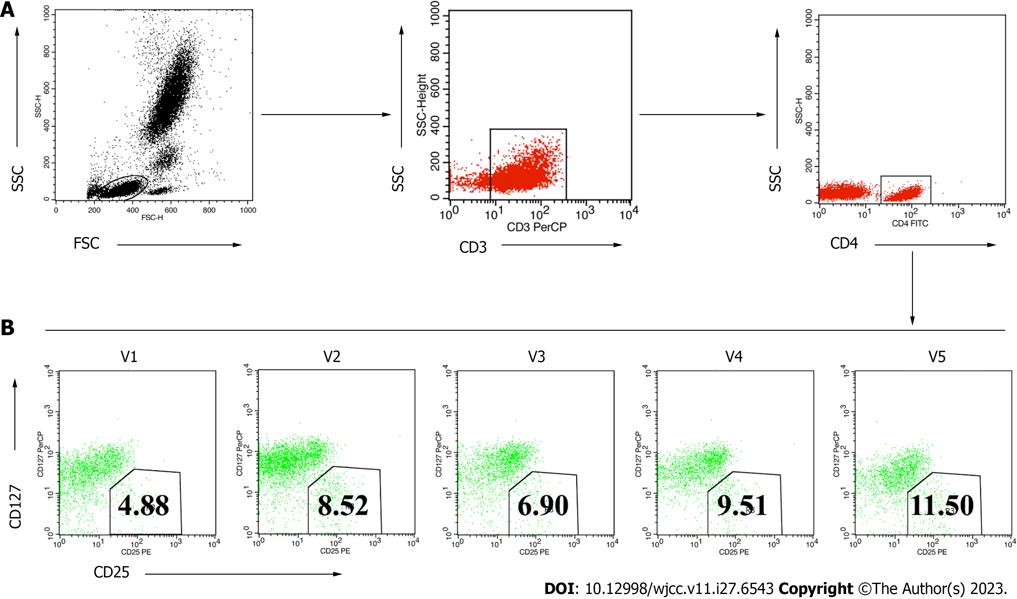
Figure 4 Fluorescence activated cell sorting results of circulating regulatory T cells before and after mesenchymal stromal cell-secretome treatments.
A: Fluorescence activated cell sorting gating strategy to detect regulatory T cells in peripheral blood; B: Circulating regulatory T cells were identified from the circulating CD4+ T cell population as CD4+CD25+127low/- using the gating strategy shown. FSC: Forward scatter; SSC: Side scatter; V: Follow-up visit.
- Citation: Lin FH, Yang YX, Wang YJ, Subbiah SK, Wu XY. Amniotic membrane mesenchymal stromal cell-derived secretome in the treatment of acute ischemic stroke: A case report. World J Clin Cases 2023; 11(27): 6543-6550
- URL: https://www.wjgnet.com/2307-8960/full/v11/i27/6543.htm
- DOI: https://dx.doi.org/10.12998/wjcc.v11.i27.6543


