Copyright
©The Author(s) 2023.
World J Clin Cases. Sep 26, 2023; 11(27): 6537-6542
Published online Sep 26, 2023. doi: 10.12998/wjcc.v11.i27.6537
Published online Sep 26, 2023. doi: 10.12998/wjcc.v11.i27.6537
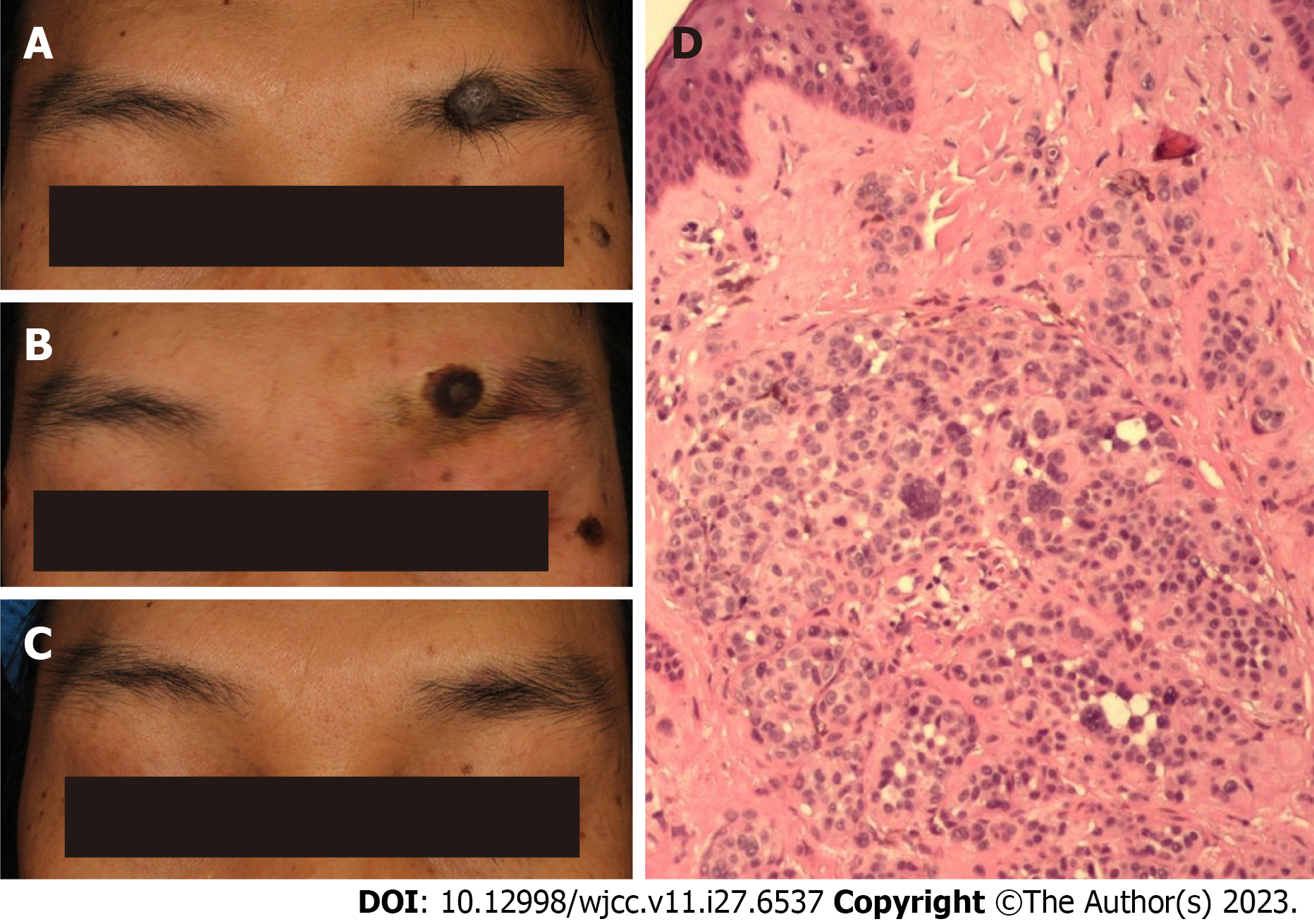
Figure 1 35-year-old male with an eyebrow intradermal nevus.
A: Before treatment, the lesion, which was located on the left brow arch, was a 13 mm round nodule with hair growth and a grey-brown appearance; B: Immediately after the surgery, there was a depressed wound on the left eyebrow arch with a dark brown scab attached, measuring approximately 10 mm; C: Thirteen months after the surgery, a light red macule measuring approximately 3 mm was observed at the site of the original skin lesion. There were no signs of depression or hypertrophic scarring, and the eyebrow was still intact; D: Under the microscope, the nevus cells in the dermis were arranged in a solid mass structure and were round, polygonal, spindle-shaped, or dendritic. The nuclei were vesicular and contained a few melanin granules.
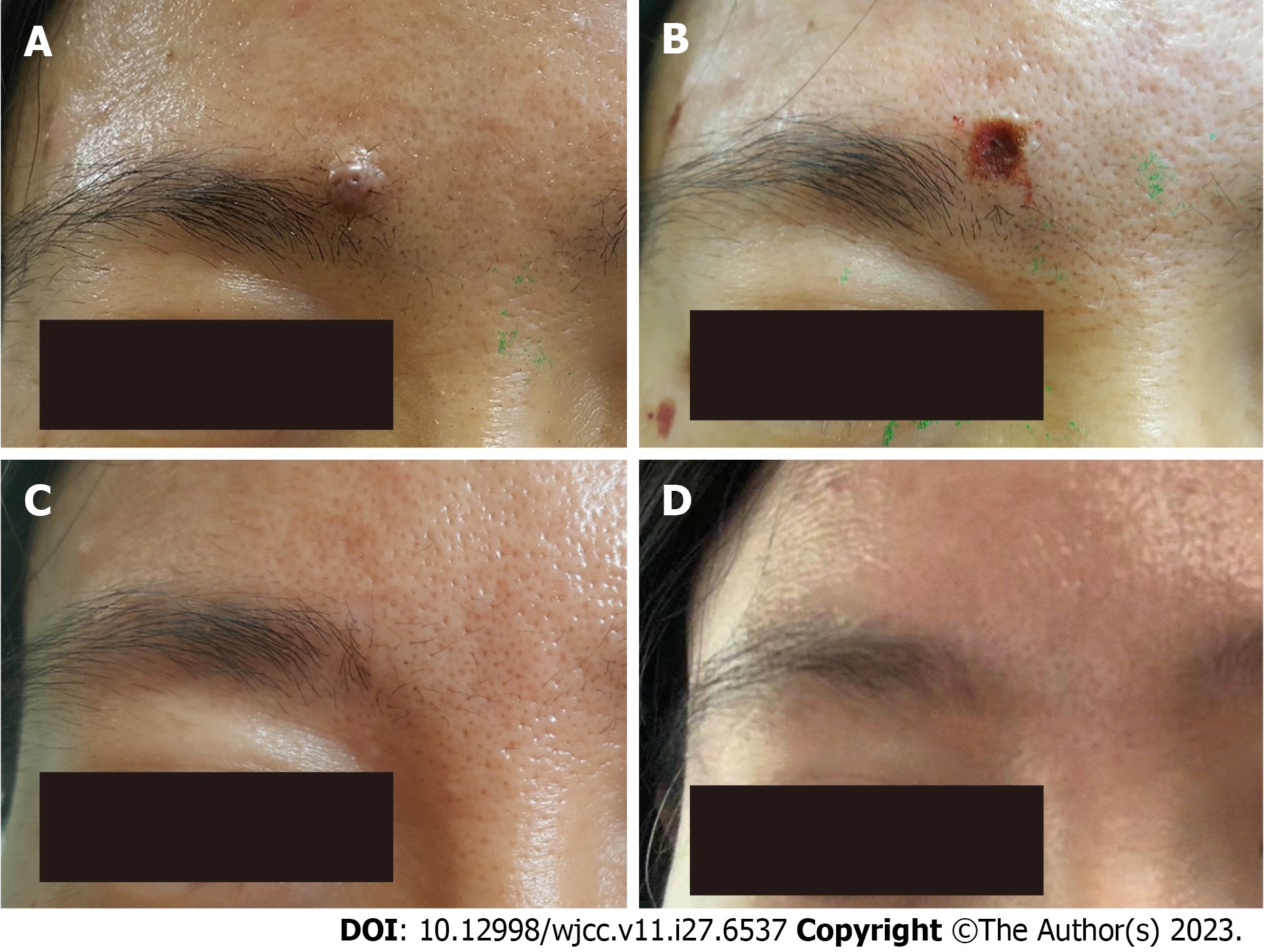
Figure 2 43-year-old female with an eyebrow intradermal nevus.
A: Before treatment, there was a skin-coloured hemispherical papule with a few black spots and hair growth on the right brow, measuring approximately 8 mm in diameter; B: Immediately after the surgery, there was a depressed, dark brown, crusted wound on the right eyebrow, measuring approximately 9 mm; C: Approximately 5 mo after surgery, an inconspicuous 2 mm circular, light-coloured rash, without depressed scars, was seen at the site of the original lesion; D: Nearly 4 years post-surgery, the skin at the site of the operation exhibited similar characteristics to the surrounding normal skin, with no signs of pigmentated or depigmented spots, as well as no evidence of sagging or hypertrophic scarring.
- Citation: Liu C, Liang JL, Yu JL, Hu Q, Li CX. Successful treatment of eyebrow intradermal nevi by shearing combined with electrocautery and curettage: Two case reports. World J Clin Cases 2023; 11(27): 6537-6542
- URL: https://www.wjgnet.com/2307-8960/full/v11/i27/6537.htm
- DOI: https://dx.doi.org/10.12998/wjcc.v11.i27.6537


