Copyright
©The Author(s) 2023.
World J Clin Cases. Sep 26, 2023; 11(27): 6483-6490
Published online Sep 26, 2023. doi: 10.12998/wjcc.v11.i27.6483
Published online Sep 26, 2023. doi: 10.12998/wjcc.v11.i27.6483
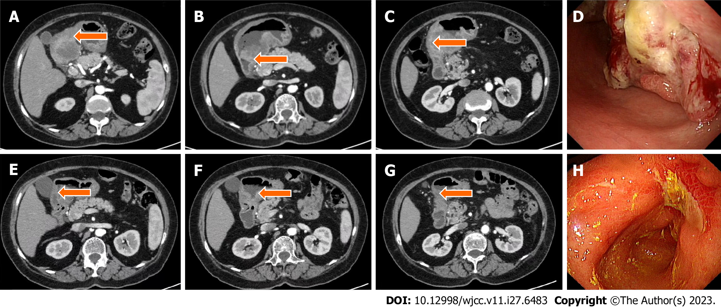
Figure 1 Computed tomography and gastroscopy images before and after treatment.
A–C: Computed tomography (CT) images of primary lesions before immunotherapy combined with chemotherapy; E–G: CT image of the primary lesion significantly reduced after immunotherapy combined with chemotherapy; D: Gastroscopy image before immunotherapy combined with chemotherapy; H: After immunotherapy combined with chemotherapy, the antral mass was significantly reduced by gastroscopy.
Figure 2 Pathological section.
A: Hematoxylin and eosin staining of tumor specimens before surgery; B: Postoperative pathology showed no residual tumor cells, and a complete pathological response was achieved; C: Needle biopsy of liver metastases revealed adenocarcinoma. A–C: Scale bar: 50 μm. Magnification: 200×.
Figure 3 Detection of mismatch repair protein.
A: MLH1 was detected by immunohistochemistry; B: MSH2 was detected by immunohistochemistry; C: MSH6 was detected by immunohistochemistry; D: PMS2 was detected by immunohistochemistry. A–D: Scale bar: 50 μm. Magnification: 200×.
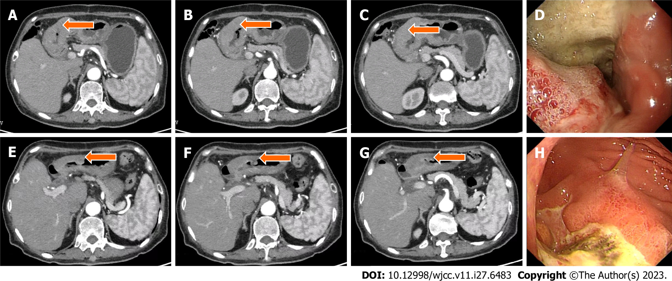
Figure 4 Computed tomography and gastroscopy images before and after treatment.
A–C: Computed tomography (CT) images of primary lesions before immunotherapy combined with chemotherapy; E–G: CT image of the primary lesion significantly reduced after immunotherapy combined with chemotherapy; D: Gastroscopy image before immunotherapy combined with chemotherapy; H: After immunotherapy combined with chemotherapy, the antral mass was significantly reduced by gastroscopy.
Figure 5 Pathological section.
A: Hematoxylin and eosin staining of tumor specimens before surgery; B: Postoperative pathology showed no residual tumor cells, and a complete pathological response was achieved; C: Examination of ascites can find heterotypic cells and consider malignancy. A–C: Scale bar: 50 μm. Magnification: 200×.
Figure 6 Detection of mismatch repair protein.
A: MLH1 was detected by immunohistochemistry; B: MSH2 was detected by immunohistochemistry; C: MSH6 was detected by immunohistochemistry; D: PMS2 was detected by immunohistochemistry. A–D: Scale bar: 50 μm. Magnification: 200×.
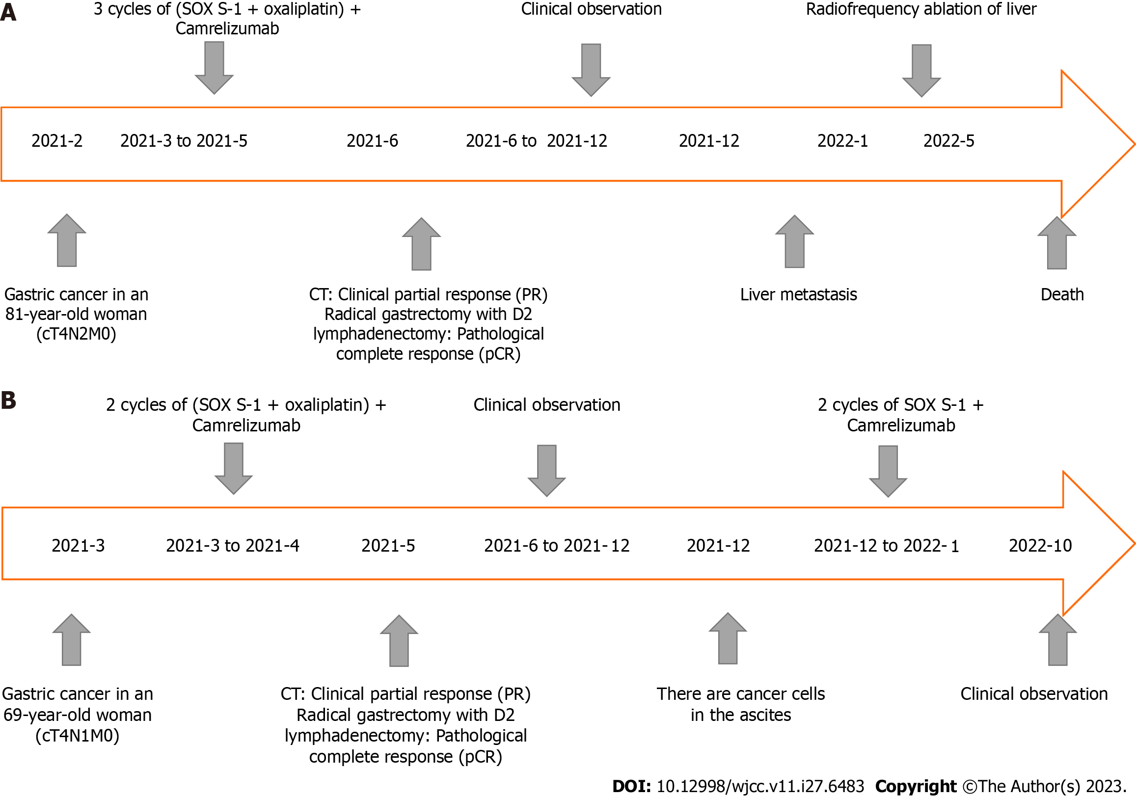
Figure 7 Timeline of diagnosis and treatment.
A: Case 1; B: Case 2.
- Citation: Xing Y, Zhang ZL, Ding ZY, Song WL, Li T. Tumor recurrence after pathological complete response in locally advanced gastric cancer after neoadjuvant therapy: Two case reports. World J Clin Cases 2023; 11(27): 6483-6490
- URL: https://www.wjgnet.com/2307-8960/full/v11/i27/6483.htm
- DOI: https://dx.doi.org/10.12998/wjcc.v11.i27.6483


