Copyright
©The Author(s) 2023.
World J Clin Cases. Sep 26, 2023; 11(27): 6476-6482
Published online Sep 26, 2023. doi: 10.12998/wjcc.v11.i27.6476
Published online Sep 26, 2023. doi: 10.12998/wjcc.v11.i27.6476
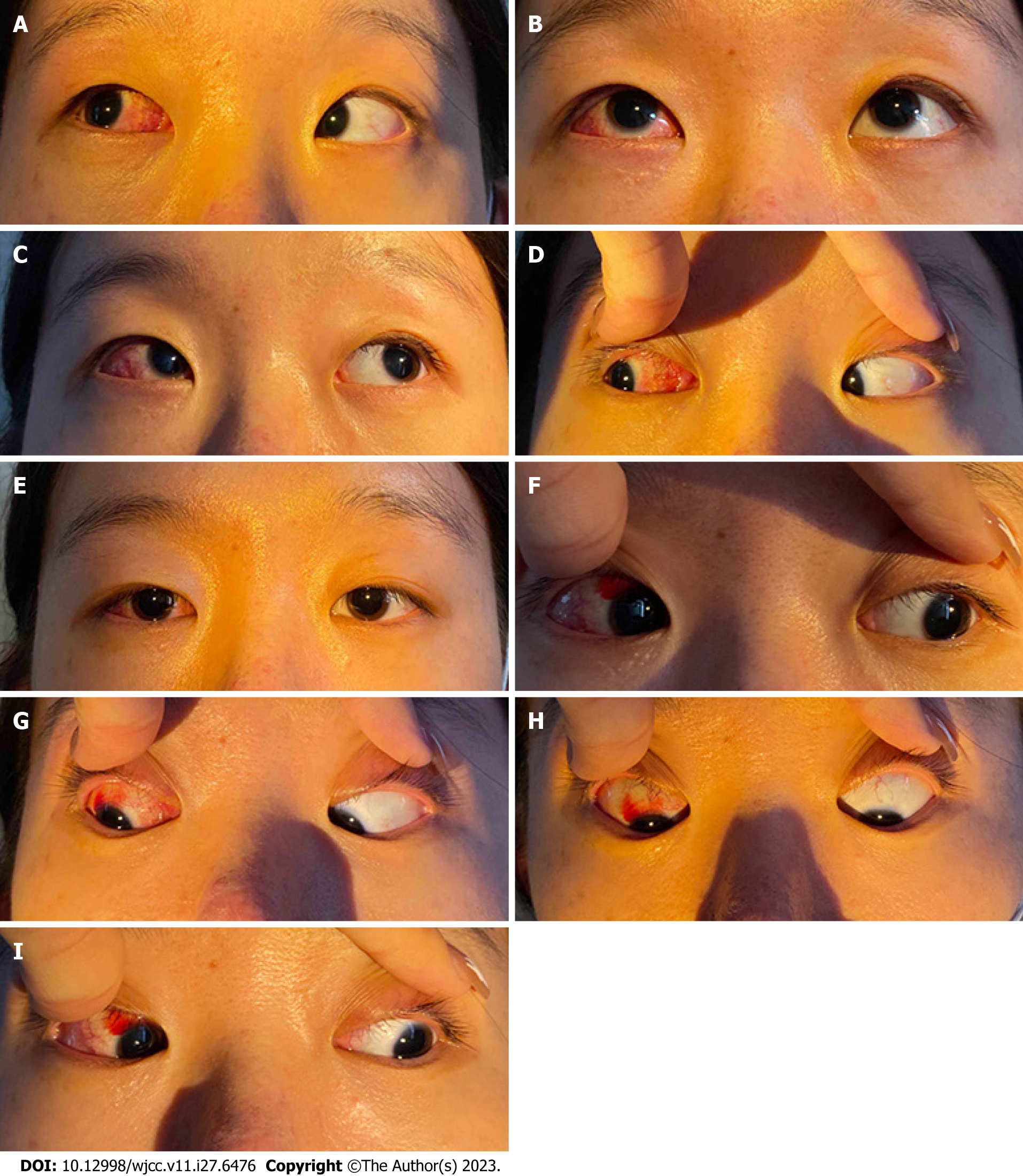
Figure 1 Preoperative nine-gaze image.
A: Normal eye movement in gaze up-and-right; B: Normal eye movement in upgaze; C: Overaction of the inferior oblique muscle in the right eye; D: Normal eye movement in right gaze; E: Esotropia of the right eye in the primary position; F: Overaction of the inferior oblique muscle in the right eye; G: Normal eye movement in gaze down-and-right; H: Normal eye movement in downgaze; I: Underaction of the superior oblique muscle in the right eye.
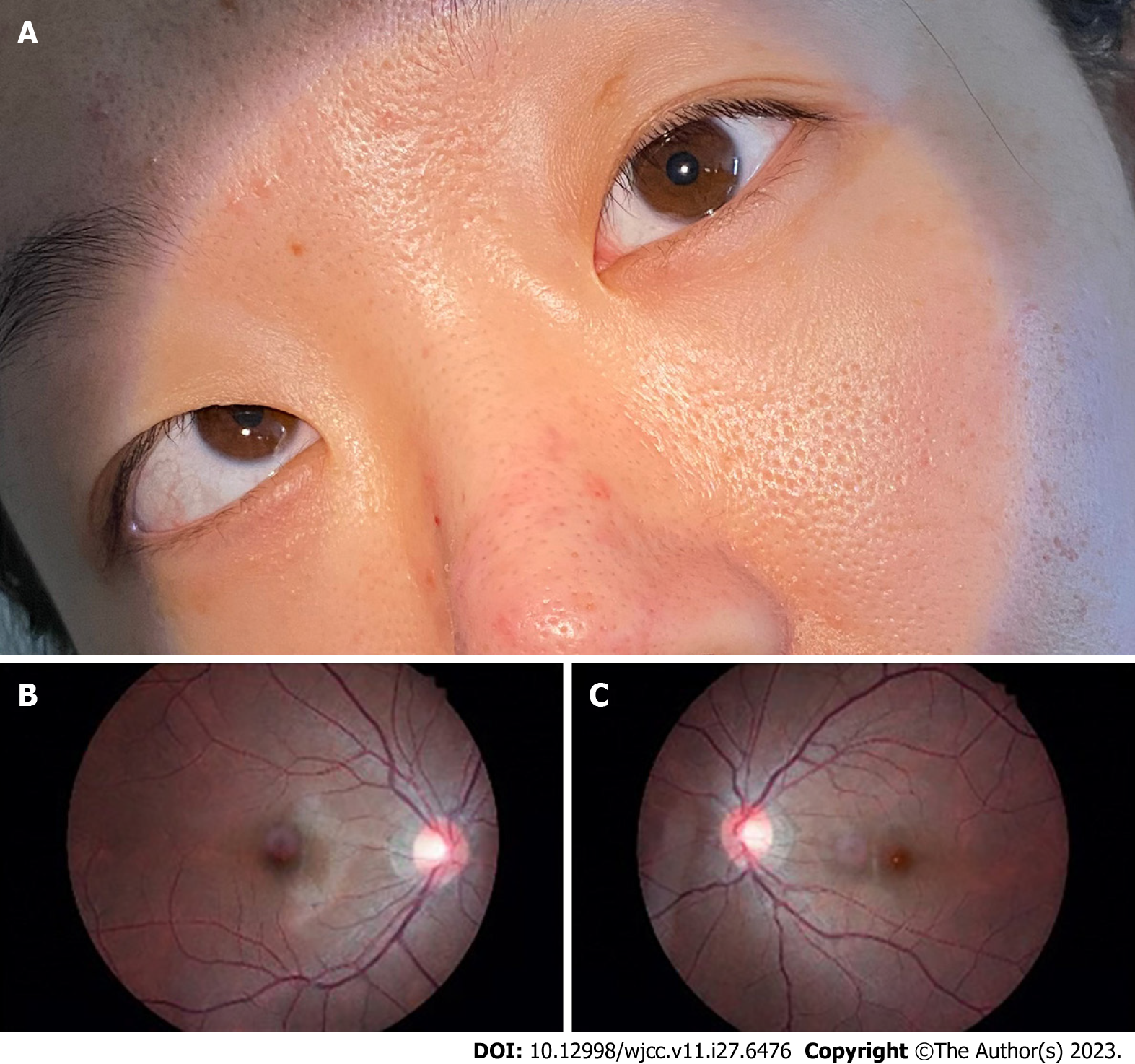
Figure 2 Preoperative Bielschowsky head tilt test and fundus photography.
A: Bielschowsky head tilt test showing right hypertropia worse with right head tilt; B and C: Preoperative fundus photography showing a normal fovea-disc relative position in the right (B) and left (C) eyes.
Figure 3 Postoperative nine-gaze image 1 wk after surgery.
A: Normal eye movement in gaze up-and-right; B: Normal eye movement in upgaze; C: Improved inferior oblique overaction in the right eye; D: Normal eye movement in right gaze; E: Orthophoria in the primary position; F: Improved inferior oblique overaction in the right eye; G: Normal eye movement in gaze down-and-right; H: Normal eye movement in downgaze. I: Improved superior oblique underaction in the right eye.
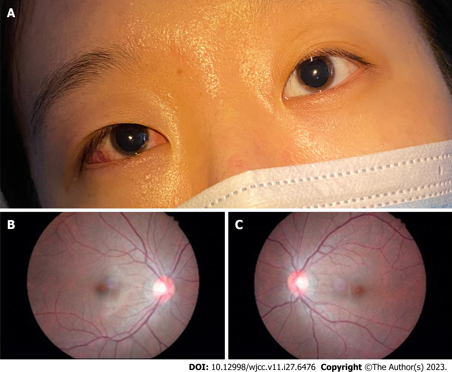
Figure 4 Postoperative Bielschowsky head tilt test and fundus photography 1 wk after surgery.
A: Bielschowsky head tilt test showing negative results; B and C: Postoperative fundus photography showing a normal fovea-disc relative position in the right (B) and left (C) eyes.
- Citation: Zhang MD, Liu XY, Sun K, Qi SN, Xu CL. Acute acquired concomitant esotropia with congenital paralytic strabismus: A case report. World J Clin Cases 2023; 11(27): 6476-6482
- URL: https://www.wjgnet.com/2307-8960/full/v11/i27/6476.htm
- DOI: https://dx.doi.org/10.12998/wjcc.v11.i27.6476


