Copyright
©The Author(s) 2023.
World J Clin Cases. Sep 26, 2023; 11(27): 6424-6430
Published online Sep 26, 2023. doi: 10.12998/wjcc.v11.i27.6424
Published online Sep 26, 2023. doi: 10.12998/wjcc.v11.i27.6424
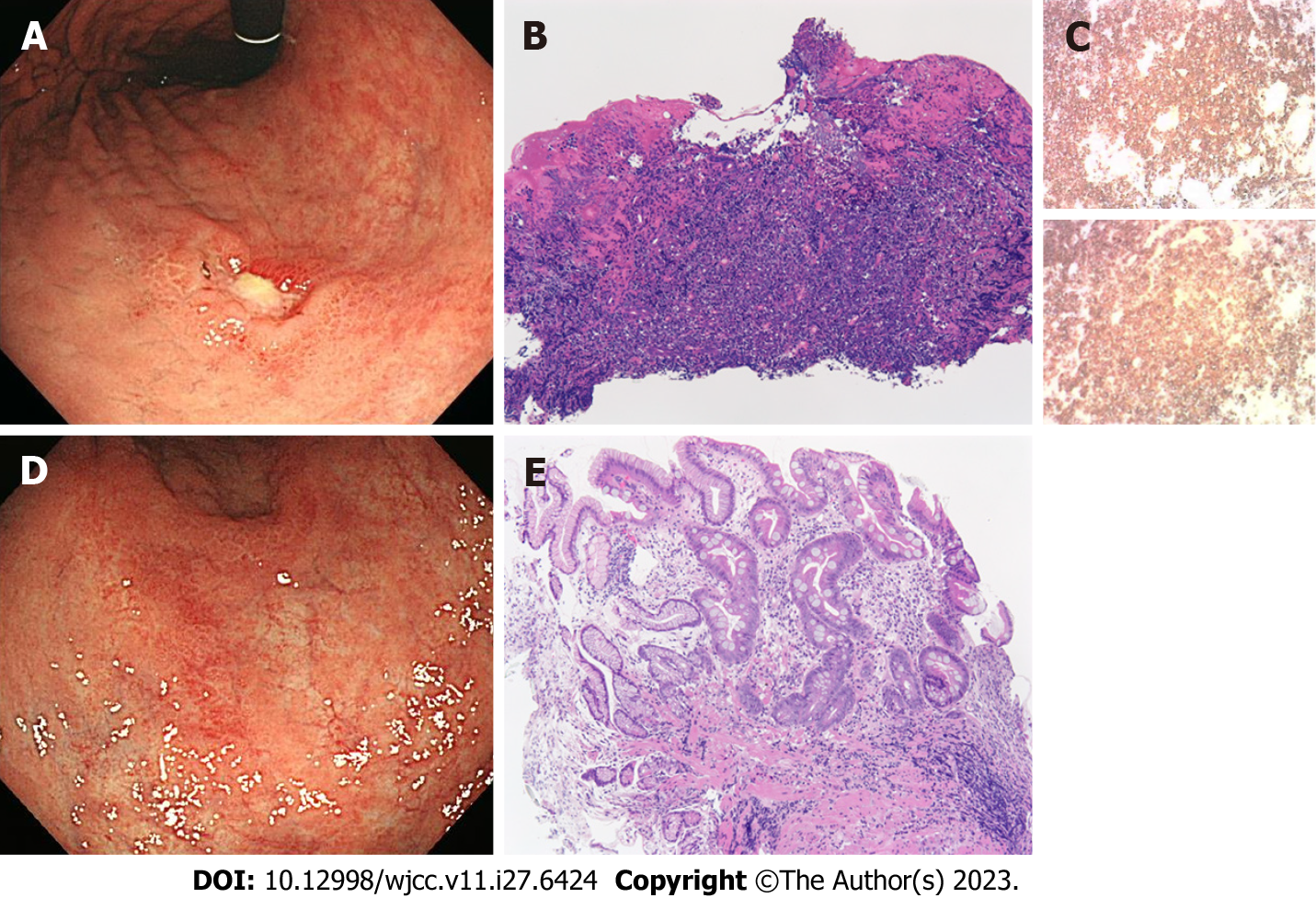
Figure 1 Case 1.
A: An ulcerated lesion was revealed in the lesser curvature of the lower corpus; B: Abnormal lymphoid cells were densely proliferating in the lamina propria; C: The upper panel shows CD20 immunostaining, and the lower panel shows CD79a immunostaining. Both were clearly positive; D: Seven months after Helicobacter pylori eradication, the ulcerated lesion regressed; E: Scattered infiltration of small lymphocytes in the lamina propria was observed. A and D: Endoscopic findings of the stomach; B, C, and E: Microscopic findings (B and E; hematoxylin eosin staining, scale bar = 100 μm).
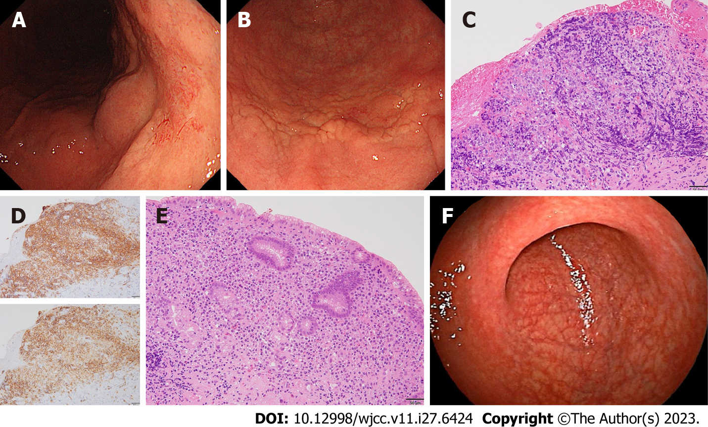
Figure 2 Case 2.
A: The surface layer of the mucosa on the posterior wall side changed to reddish; B: A subtle ridged change in the mucosa was observed; C: Neoplastic lymphoid cells were densely proliferating in the lamina propria; D: The upper panel shows CD20 immunostaining, and the lower panel shows CD79a immunostaining. Both were clearly positive; E: Atypical lymphoid cells were proliferating in the lamina propria; F: Fourteen months after Helicobacter pylori eradication, a white scar was observed on the posterior wall where diffuse large B-cell lymphoma was present. A, B, and F: Endoscopic findings of the stomach; C-E: Microscopic findings (C and E; hematoxylin hosin staining, scale bar = 100 μm).
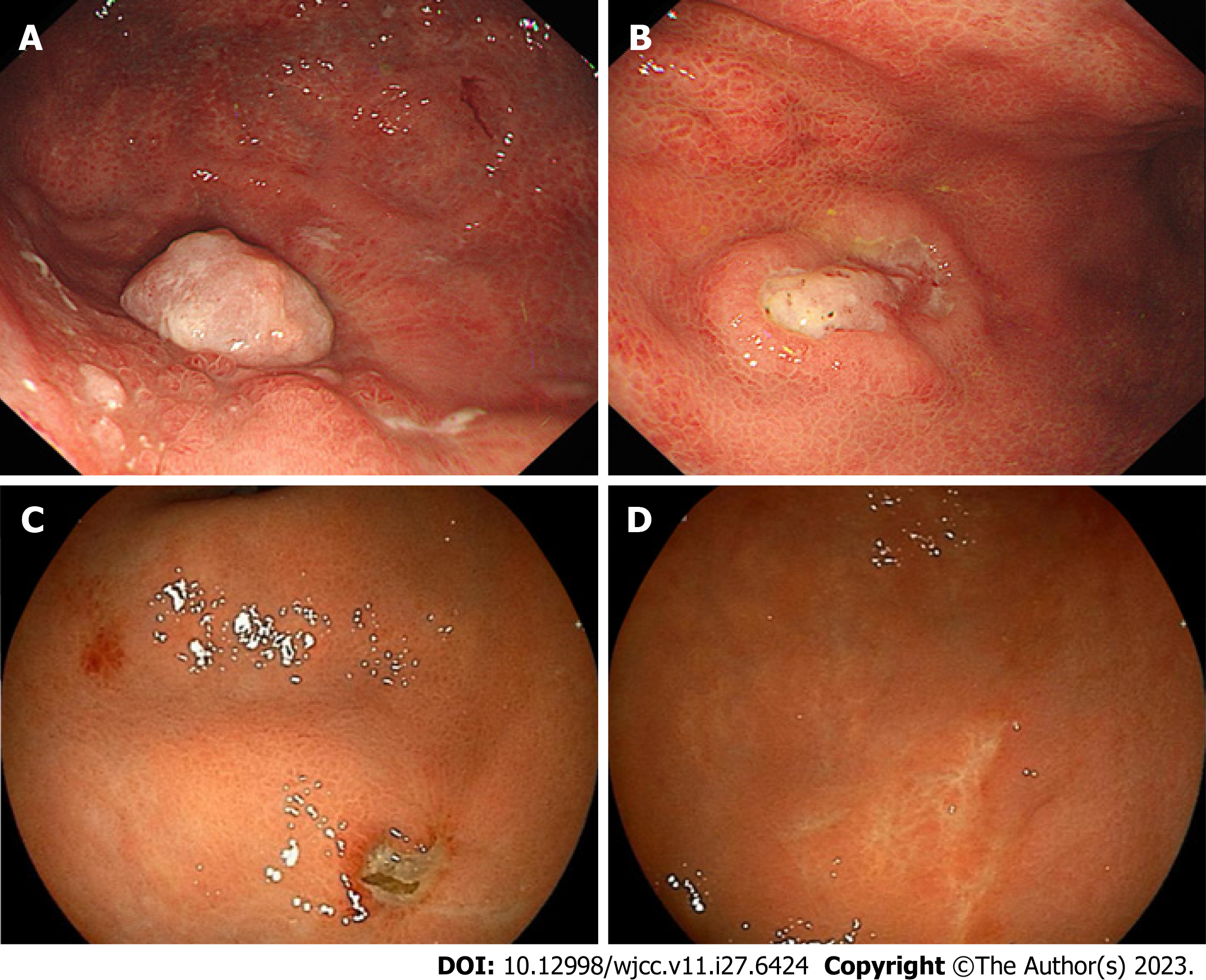
Figure 3 Case 3.
Endoscopic findings of the stomach. A: A polypoid-like elevated lesion was found in the fundus; B: An ulcerative lesion with peripheral ridges was found in the greater curvature of the corpus; C and D: Three months after Helicobacter pylori eradication, the elevated lesion in the fundus was ulcerated (C), and the ulcerative lesions in the corpus became scarred (D).
- Citation: Saito M, Mori A, Kajikawa S, Yokoyama E, Kanaya M, Izumiyama K, Morioka M, Kondo T, Tanei ZI, Shimizu A. Helicobacter pylori eradication treatment for primary gastric diffuse large B-cell lymphoma: A single-center analysis. World J Clin Cases 2023; 11(27): 6424-6430
- URL: https://www.wjgnet.com/2307-8960/full/v11/i27/6424.htm
- DOI: https://dx.doi.org/10.12998/wjcc.v11.i27.6424


