Copyright
©The Author(s) 2023.
World J Clin Cases. Sep 26, 2023; 11(27): 6374-6382
Published online Sep 26, 2023. doi: 10.12998/wjcc.v11.i27.6374
Published online Sep 26, 2023. doi: 10.12998/wjcc.v11.i27.6374
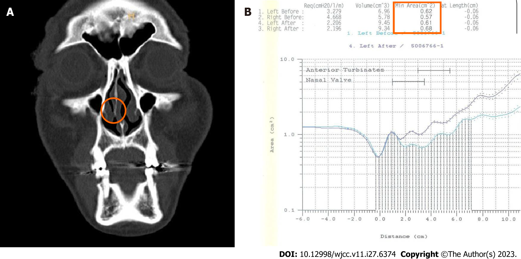
Figure 1 Preoperative facial bone computed tomography and preoperative and postoperative acoustic rhinometry.
A: Preoperative facial bone computed tomography. Left side wall fracture with septal deviation and the right-side inferior turbinate was narrowing by septal deviation; B: Preoperative and postoperative acoustic rhinometry. Change of right nasal cavity minimal cross-sectional area after closed reduction with out-fracture of the inferior turbinate.
Figure 2 Intraoperative findings.
A: A medium-sized speculum is inserted just beyond the nostril rim, with care being exercised to avoid tearing the vestibular mucosa; B: The blades are spread. Usually, a crunching sound is heard as the bony septum centralizes and the turbinate in-fractures.
Figure 3 Photographs showing the surgical methods.
A: Inferior turbinate out-fracture method; B: In-fracturing of the inferior turbinate.
Figure 4 Graphs showing the improvement according to lateral or frontal impact group.
A: In the lateral impact group, 26 patients experienced an improvement in their nasal obstruction symptoms 2 wk after the operation. An additional 6 patients also showed an improvement 4 wk later; B: In the frontal impact group, 14 patients showed an improvement in nasal obstruction symptoms 2 wk after the operation. After 4 wk, all patients reported being satisfied with the results.
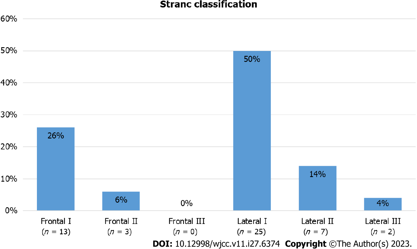
Figure 5 Nasal bone fracture type according to Stranc classification.
Figure 6 Mean minimal cross-sectional area for fracture group; frontal impact, lateral impact, total.
MCA: Minimal cross-sectional area.
- Citation: Kim SY, Nam HJ, Byeon JY, Choi HJ. Effectiveness of out-fracture of the inferior turbinate with reduction nasal bone fracture. World J Clin Cases 2023; 11(27): 6374-6382
- URL: https://www.wjgnet.com/2307-8960/full/v11/i27/6374.htm
- DOI: https://dx.doi.org/10.12998/wjcc.v11.i27.6374


