Copyright
©The Author(s) 2023.
World J Clin Cases. Sep 16, 2023; 11(26): 6304-6310
Published online Sep 16, 2023. doi: 10.12998/wjcc.v11.i26.6304
Published online Sep 16, 2023. doi: 10.12998/wjcc.v11.i26.6304
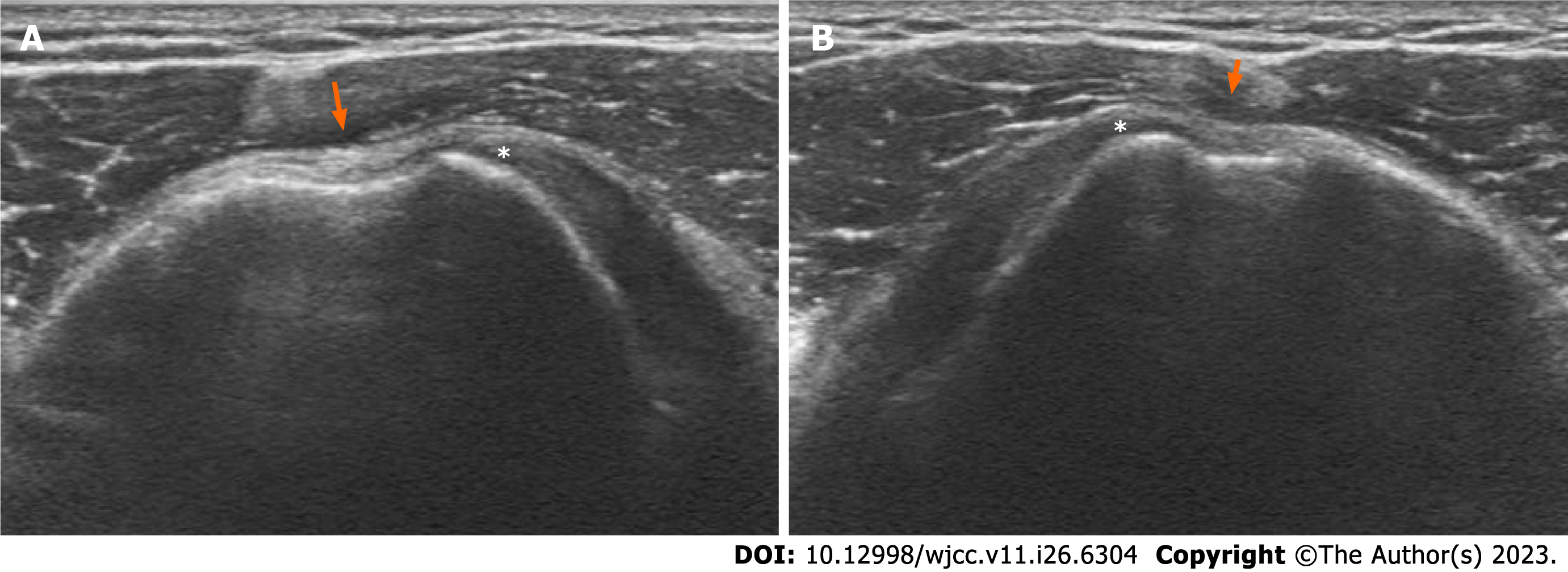
Figure 1 Ultrasonography.
A: Left shoulder; B: Right shoulder. Intact subscapularis tendon (asterisk) and empty bicipital groove with dysplasia (arrow).
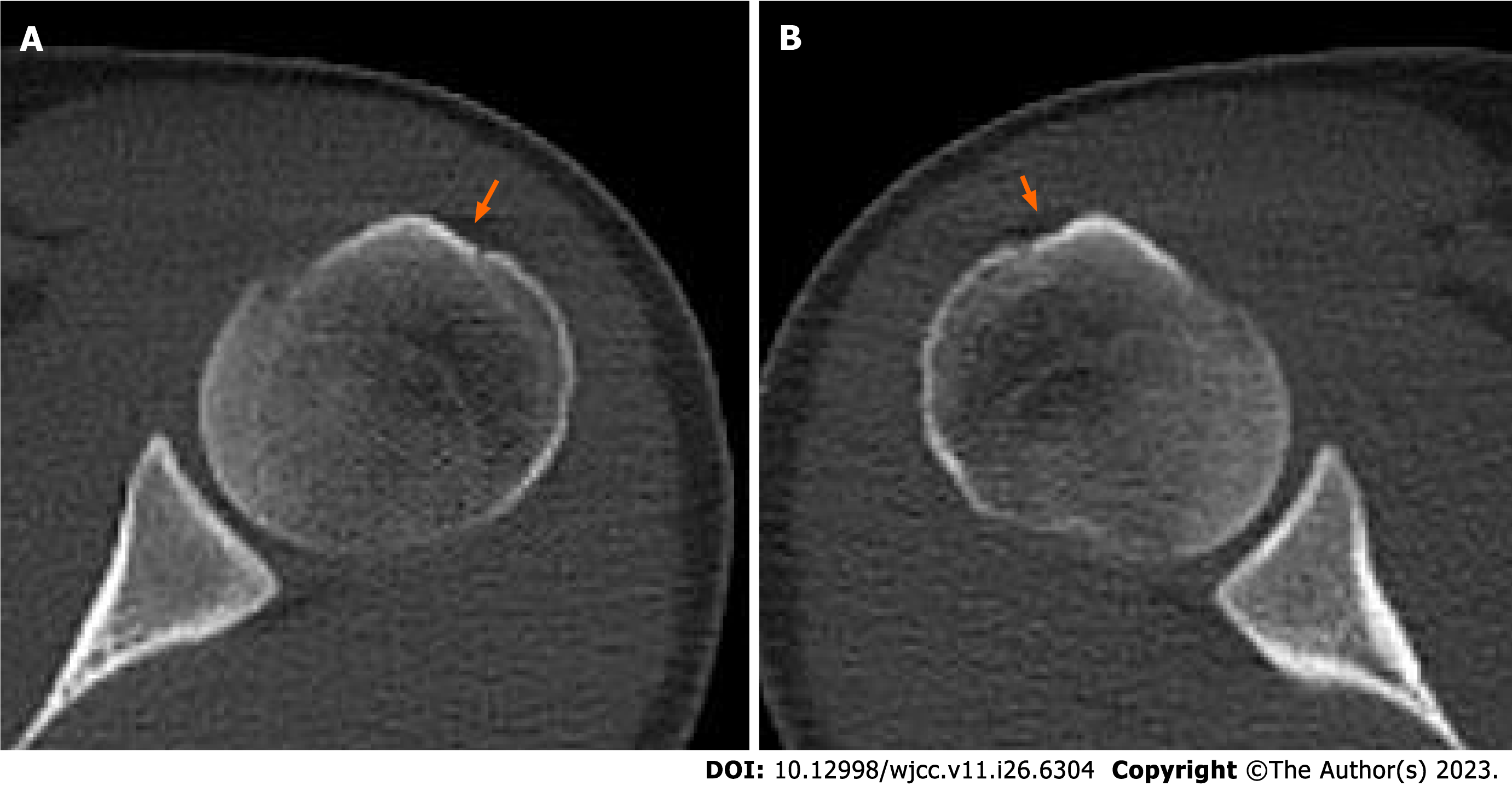
Figure 2 Computed tomography scan showing bicipital groove dysplasia.
A: Left shoulder; B: Right shoulder.
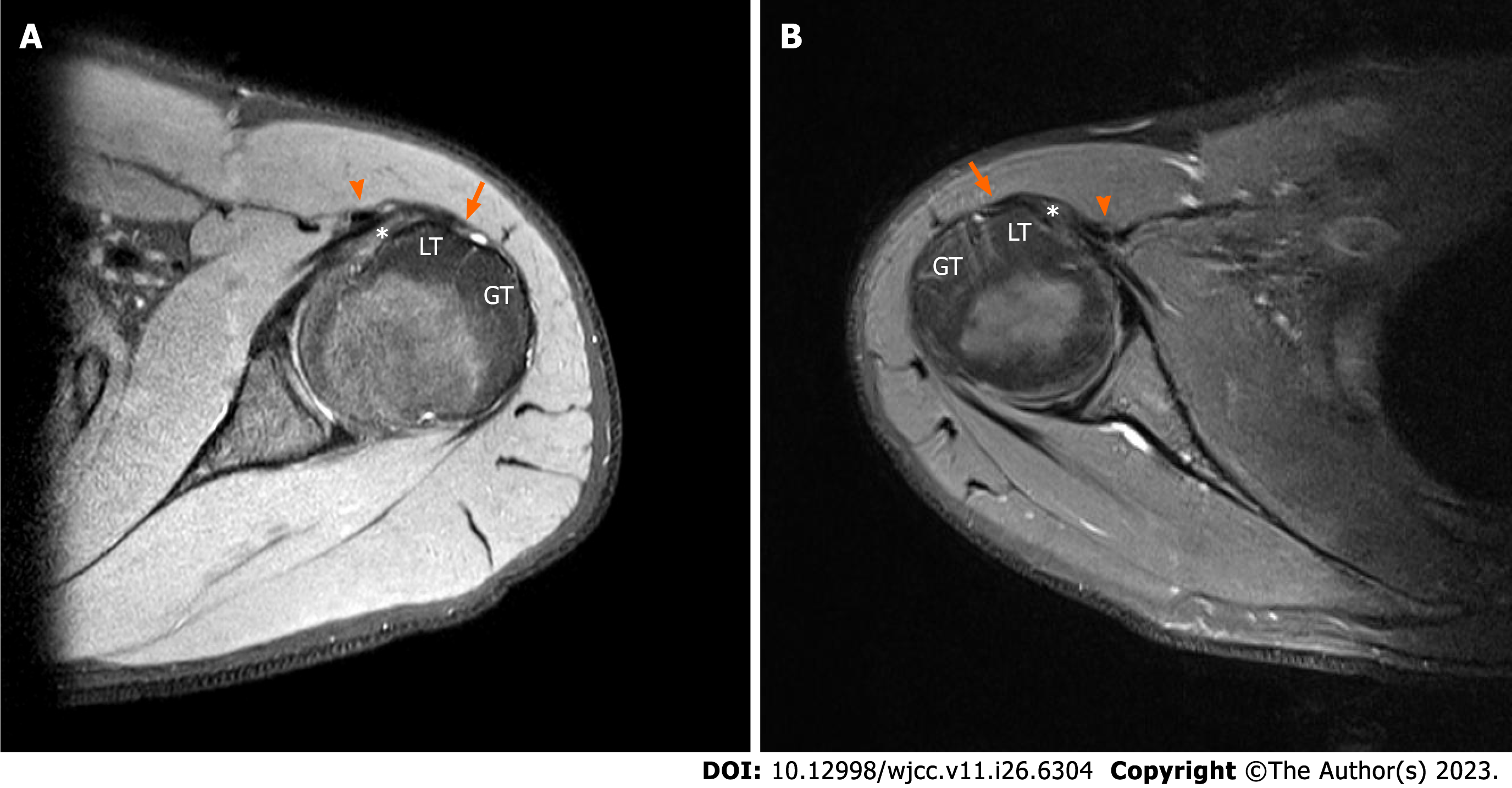
Figure 3 Magnetic resonance imaging axial T2w.
A: Left shoulder; B: Right shoulder. Dislocation of the long head of biceps tendon (triangle) over the intact subscapularis tendon (asterisk). Flattening of bicipital groove is remarkable (arrow). Long head of biceps tendon distance of left shoulder: 17.9 mm; right shoulder: 20.9 mm. LT: Lesser tuberosity; GT: Greater tuberosity.
Figure 4 Arthroscopic photo from the posterior portal.
A: Left shoulder; B: Right shoulder. Dislocation of the biceps tendon (triangle), fraying of biceps pulley (arrow), and intact subscapularis tendon (asterisk). HH: Humeral head.
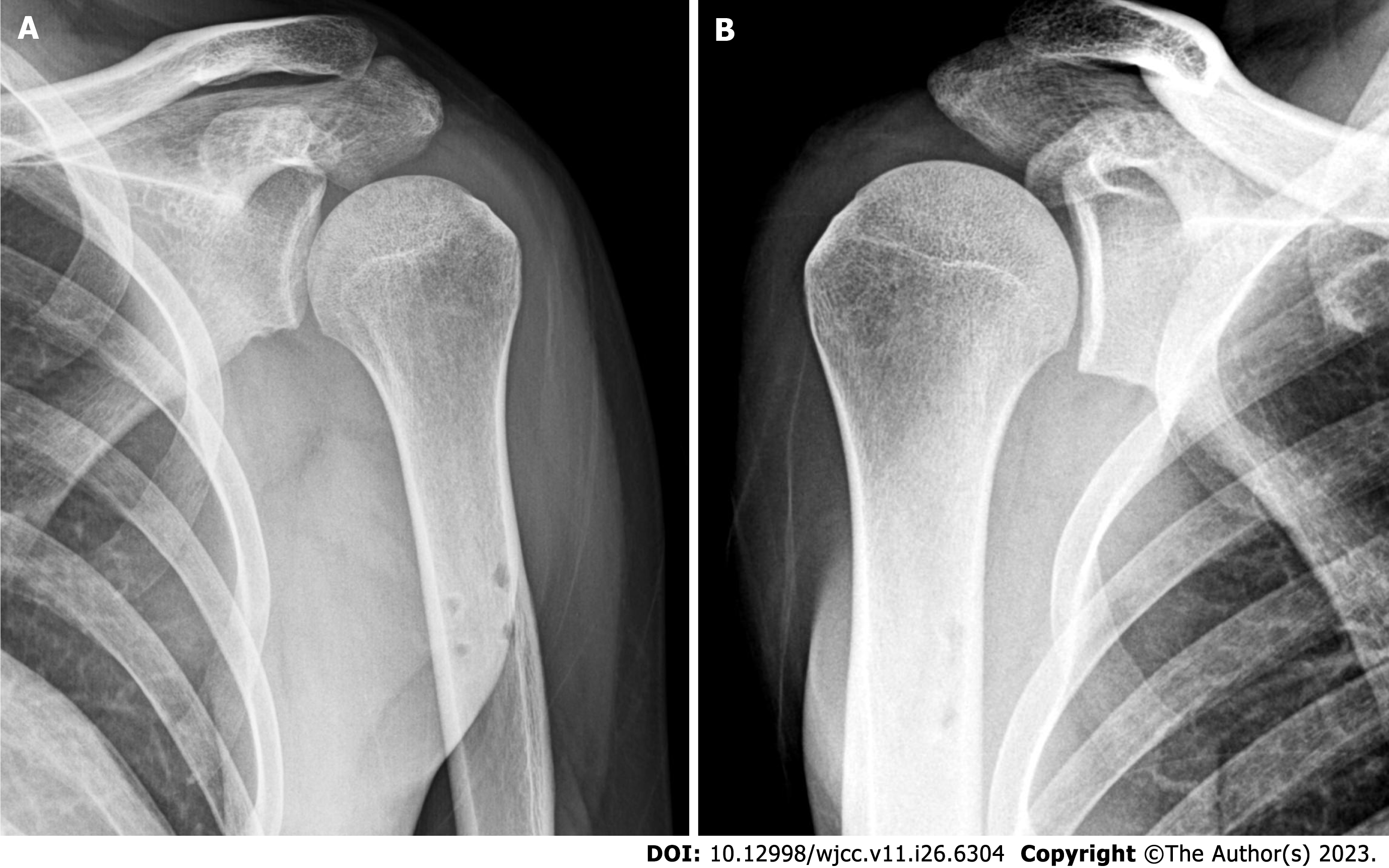
Figure 5 Postoperative radiographs after subpectoral tenodesis.
A: Left shoulder; B: Right shoulder.
- Citation: Sohn HJ, Cho CH, Kim DH. Bilateral dislocation of the long head of biceps tendon with intact rotator cuff tendon: A case report. World J Clin Cases 2023; 11(26): 6304-6310
- URL: https://www.wjgnet.com/2307-8960/full/v11/i26/6304.htm
- DOI: https://dx.doi.org/10.12998/wjcc.v11.i26.6304


