Copyright
©The Author(s) 2023.
World J Clin Cases. Sep 16, 2023; 11(26): 6298-6303
Published online Sep 16, 2023. doi: 10.12998/wjcc.v11.i26.6298
Published online Sep 16, 2023. doi: 10.12998/wjcc.v11.i26.6298
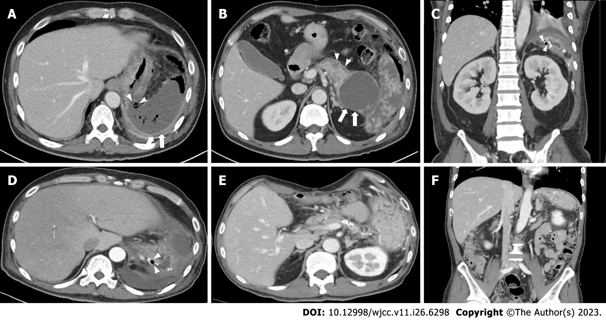
Figure 1 Abdominal contrast-enhanced computed tomography images in a 45-year-old man.
A: Adjacent to the huge walled-off necrosis, there is a wall defect, demonstrating perforation of the stomach fundus (arrowheads) and splenic infarction (white arrow); B: Contrast-enhanced computed tomography (CT) scanning demonstrated the huge walled-off necrosis at the intra- (arrowheads) and extrapancreatic areas (white arrows); C: Axial view of contrast-enhanced CT image; D: Coronary view of portal venous phase CT on postoperative day 18 shows significant wall defect on previous staple line (arrowheads); E and F: Axial and coronary views of portal venous phase CT at the 3-mo follow-up. CT images show improved process of loculated fluid collection with air bubble at pancreatic bed and left subphrenic space.
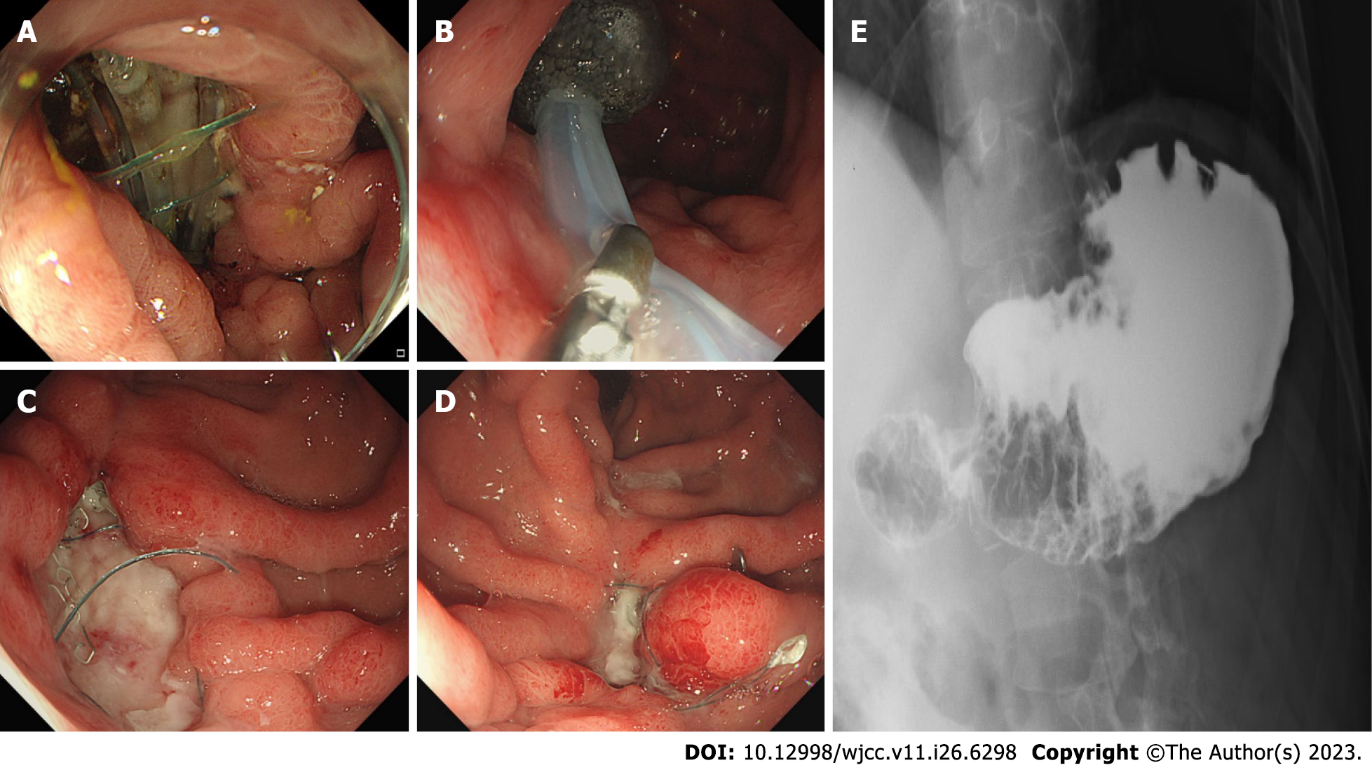
Figure 2 Treatment.
A: 45-year-old man is diagnosed with a 3-cm gastric perforation at the anastomosis site on postoperative day 18; B: A polyurethane sponge is inserted into the cavity of the anastomotic leak with nasogastric continuous suction; C: The perforation site is downsized with granulation tissue during the fourth endoscopic vacuum-assisted closure (EVAC); D: The cavity is closed after seven EVAC procedures; E: Follow-up upper gastrointestinal radiography shows no contrast leakage from the stomach.
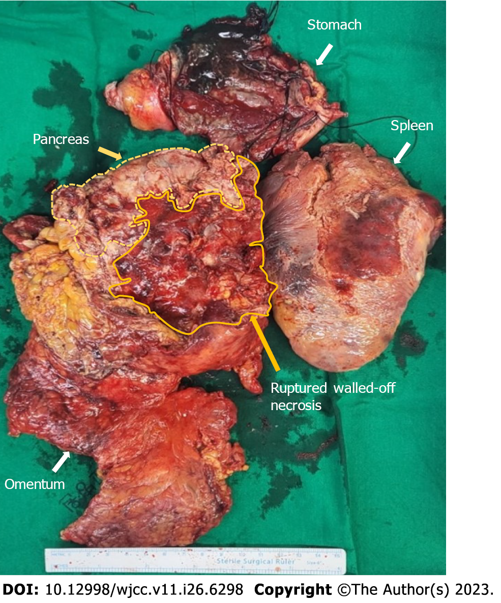
Figure 3 Surgical specimen after distal pancreatectomy, splenectomy, and gastric wedge resection.
Note that the pancreatic walled-off necrosis is ruptured during operation. Each specimen is resected separately.
- Citation: Noh BG, Yoon M, Park YM, Seo HI, Kim S, Hong SB, Park JK, Lee MW. Successful resolution of gastric perforation caused by a severe complication of pancreatic walled-off necrosis: A case report. World J Clin Cases 2023; 11(26): 6298-6303
- URL: https://www.wjgnet.com/2307-8960/full/v11/i26/6298.htm
- DOI: https://dx.doi.org/10.12998/wjcc.v11.i26.6298


