Copyright
©The Author(s) 2023.
World J Clin Cases. Sep 16, 2023; 11(26): 6262-6267
Published online Sep 16, 2023. doi: 10.12998/wjcc.v11.i26.6262
Published online Sep 16, 2023. doi: 10.12998/wjcc.v11.i26.6262
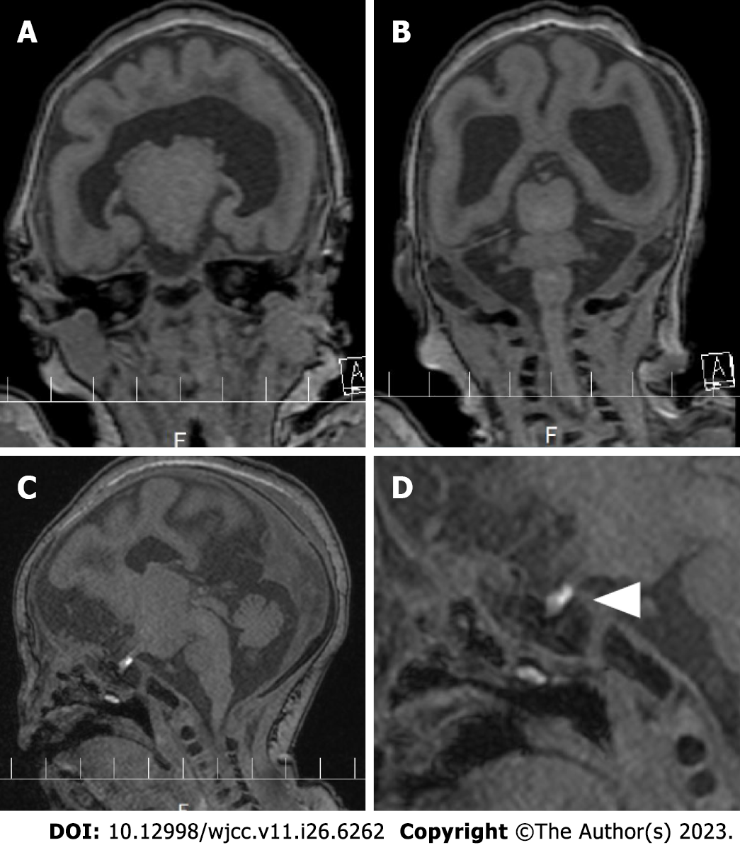
Figure 1 Head magnetic resonance imaging.
A and B: Coronal view of a T1 weighted image. The left and right cerebral hemispheres are incomplete, and the corpus callosum is absent, and there is a semilobar encephalocele; C and D: Parasagittal view of a T1 weighted image. The triangle indicates an ectopic posterior pituitary stalk.
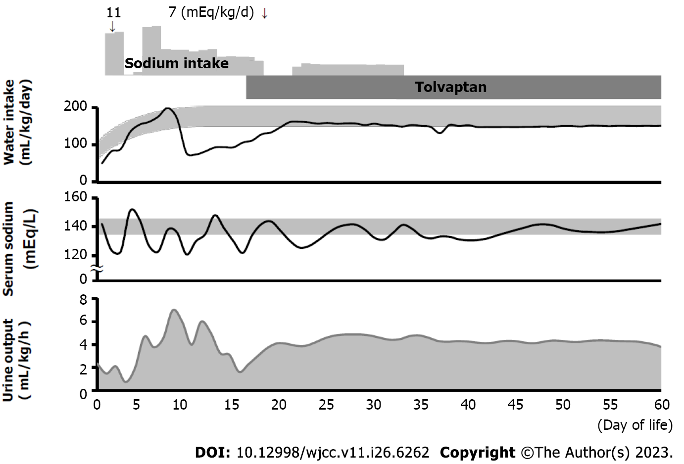
Figure 2 Clinical course of the patient.
Oral administration of sodium and tolvaptan, water intake, serum sodium level, and urine output are shown. In the “Water intake” and “Serum sodium” chart, the gray band represents the general range of a healthy infant.
- Citation: Mori M, Takeshita S, Nakamura N, Mizuno Y, Tomita A, Aoyama M, Kakita H, Yamada Y. Efficacy of tolvaptan in an infant with syndrome of inappropriate antidiuretic hormone secretion associated with holoprosencephaly: A case report. World J Clin Cases 2023; 11(26): 6262-6267
- URL: https://www.wjgnet.com/2307-8960/full/v11/i26/6262.htm
- DOI: https://dx.doi.org/10.12998/wjcc.v11.i26.6262


