Copyright
©The Author(s) 2023.
World J Clin Cases. Sep 16, 2023; 11(26): 6206-6212
Published online Sep 16, 2023. doi: 10.12998/wjcc.v11.i26.6206
Published online Sep 16, 2023. doi: 10.12998/wjcc.v11.i26.6206
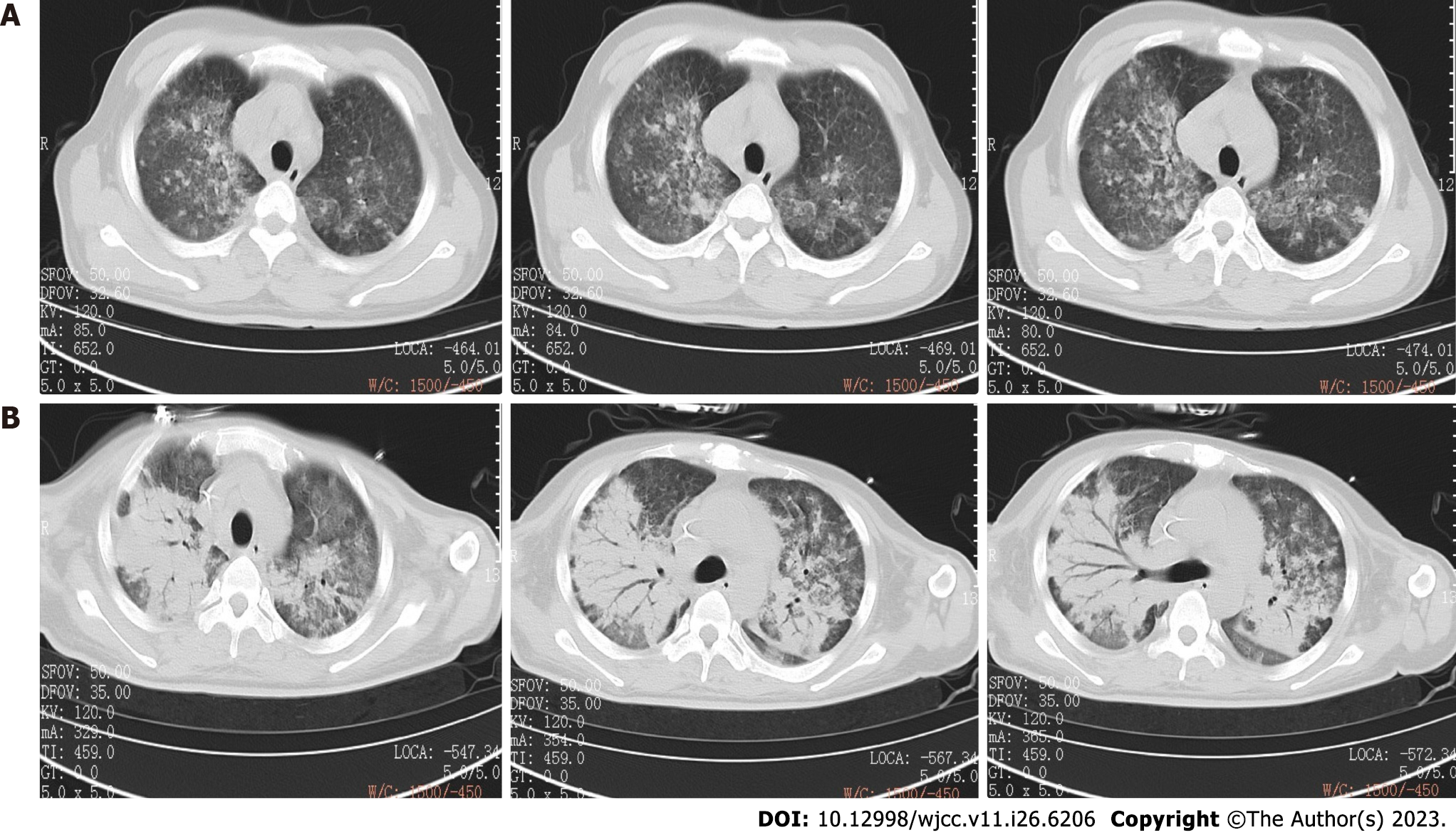
Figure 1 Computed tomography.
A: Chest computed tomography (CT) on April 11th showed that pneumonia on two sides; B: Chest CT scan on May 24th showed that pneumonia was advanced than before.
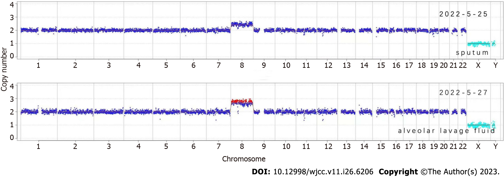
Figure 2 Copy number variation analysis of the patient’s chromosomes.
Both sputum and alveolar lavage fluid samples showed large Copy number variations on chromosome 8.
Figure 3
Bone marrow examination showed erythroid deficiency and grain maturation disorder, considering infection.
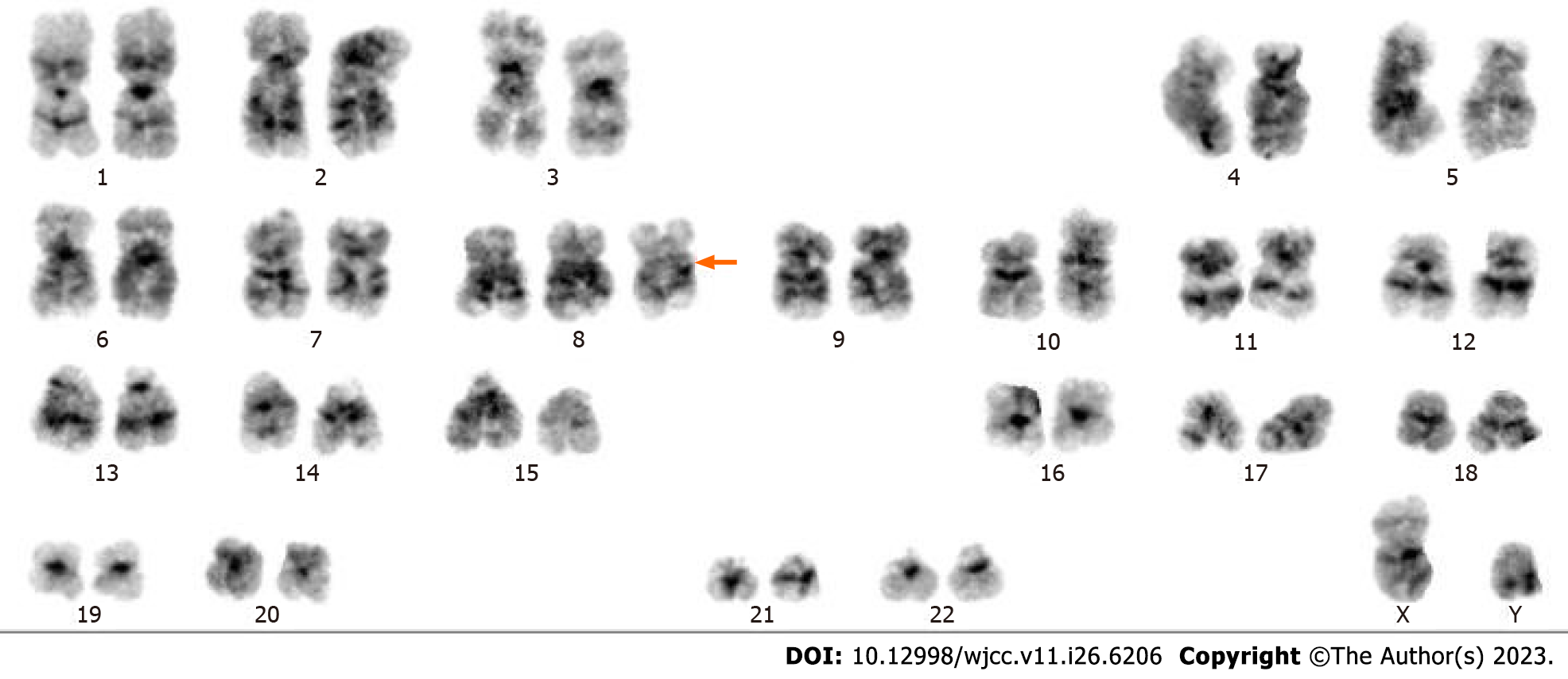
Figure 4
Chromosome analysis of bone marrow showed trisomy 8 (arrow).
- Citation: Pan FY, Fan HZ, Zhuang SH, Pan LF, Ye XH, Tong HJ. Severe inflammatory disorder in trisomy 8 without myelodysplastic syndrome and response to methylprednisolone: A case report. World J Clin Cases 2023; 11(26): 6206-6212
- URL: https://www.wjgnet.com/2307-8960/full/v11/i26/6206.htm
- DOI: https://dx.doi.org/10.12998/wjcc.v11.i26.6206


