Copyright
©The Author(s) 2023.
World J Clin Cases. Sep 16, 2023; 11(26): 6189-6193
Published online Sep 16, 2023. doi: 10.12998/wjcc.v11.i26.6189
Published online Sep 16, 2023. doi: 10.12998/wjcc.v11.i26.6189
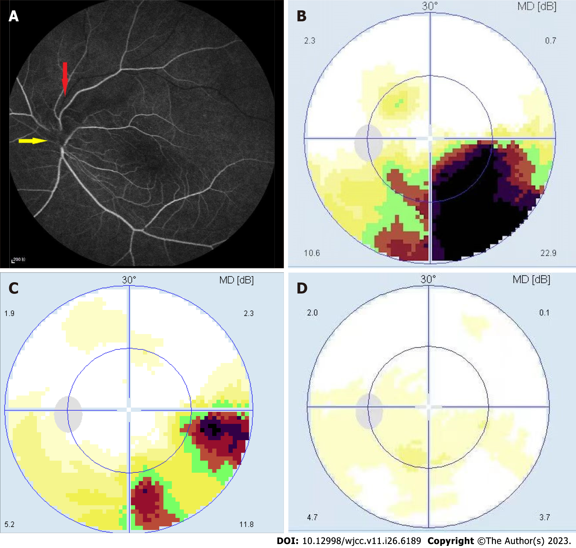
Figure 1 Fundus fluorescein angiography and visual field before and after treatment for non-arterial anterior ischemic optic neuropathy combined with retinal branch vein occlusion.
A: On this fundus fluorescein angiography image, the yellow arrow at 18.12" indicates low fluorescence above the optic disc, and the red arrow shows that the superior temporal branch vein is unfilled. Laminar flow is visible in the other branch veins; B: During the patient’s visit, a fan-shaped defect was observed in the lower visual field and was connected to the optic disc; C: The patient’s lower visual field defect improved after 2 wk of an intravenous drip; D: After 3 wk of an intravenous drip, the patient’s visual field had almost recovered completely.
- Citation: Gong HX, Xie SY. Non-arteritic anterior ischemic optic neuropathy combined with branch retinal vein obstruction: A case report. World J Clin Cases 2023; 11(26): 6189-6193
- URL: https://www.wjgnet.com/2307-8960/full/v11/i26/6189.htm
- DOI: https://dx.doi.org/10.12998/wjcc.v11.i26.6189


