Copyright
©The Author(s) 2023.
World J Clin Cases. Sep 16, 2023; 11(26): 6170-6175
Published online Sep 16, 2023. doi: 10.12998/wjcc.v11.i26.6170
Published online Sep 16, 2023. doi: 10.12998/wjcc.v11.i26.6170
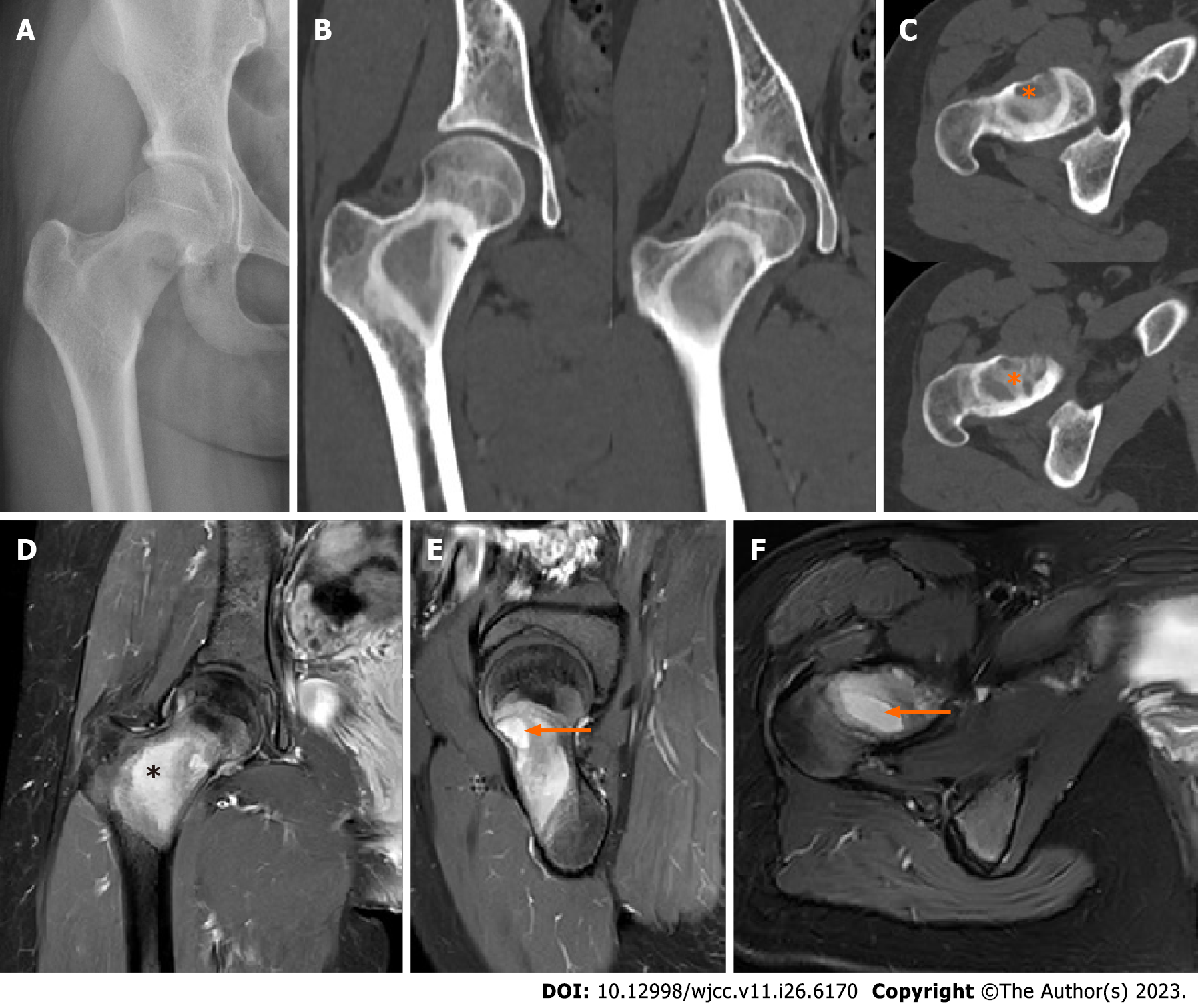
Figure 1 Preoperative radiographic images of fibrous dysplasia associated with aneurysmal-bone-cyst-like changes.
A: Preoperative anteroposterior X-ray showed ground-glass appearance with cortical scalloping and expansion of the right proximal femur and femoral neck; B and C: Preoperative computed tomography (CT). Axial CT showed multiseptated proximal femur lesion with a cyst in the right proximal femur and femoral neck (orange asterisk); D-F: Preoperative magnetic resonance imaging (MRI). Coronal and sagittal fat-suppressed, contrast-enhanced T1-weighted MRI of the right proximal femur and femoral showed multiseptated proximal femoral lesion with a dominant cystic cavity without any adjacent soft-tissue edema (black asterisk). Axial fat-suppressed, contrast-enhanced T2-weighted MRI and a sagittal fat-suppressed, contrast-enhanced T1-weighted MRI showed multiseptated proximal femoral lesion with fluid-fluid levels (orange arrows).
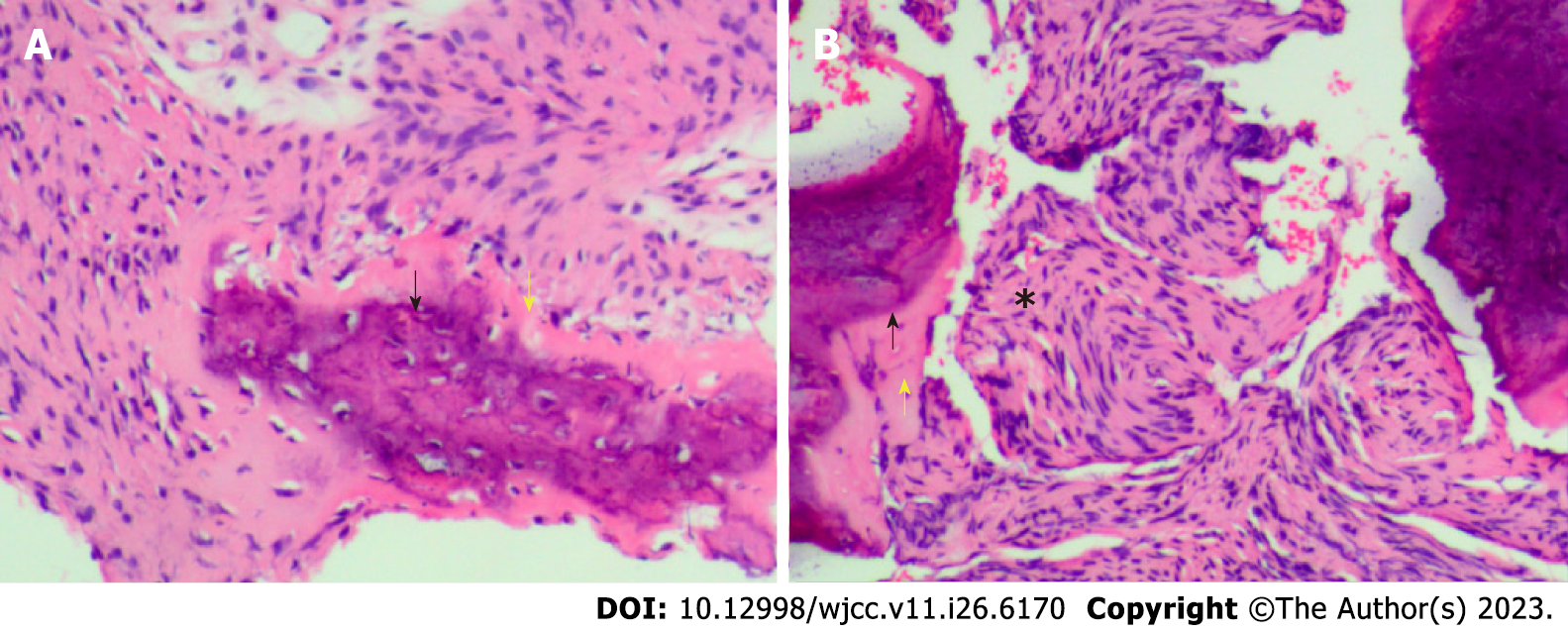
Figure 2 Pathological examination result the lesion based on preoperative puncture biopsy.
A and B: Hematoxylin and eosin staining, magnification 40’: lesions filled with irregular trabecular bone (black asterisk) and hyperplastic fibrous tissue (black arrow), with woven bone formation (yellow arrow).
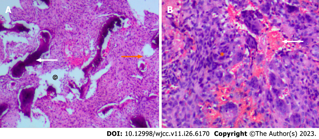
Figure 3 Postoperative pathological examination of the right proximal femur.
A and B: Hematoxylin and eosin staining, magnification 40’: Solid areas and cystic spaces (@) filled with blood (white arrow), with cellular septa (orange arrow), and hyperplastic fibrous cells (orange asterisk).
Figure 4 Radiographic images of fibrous dysplasia associated with aneurysmal-bone-cyst-like changes during follow-up.
A: Curettage and allograft associated with fixation with hip compression screws were performed; B and C: No obvious allograft absorption or any loosening of fixation or secondary fracture were observed after 3 mo, based on preoperative anteroposterior X-ray images; D-F: After 6 mo, bone fusion was observed based on computed tomography 3D assessment.
- Citation: Xie LL, Yuan X, Zhu HX, Pu D. Surgery for fibrous dysplasia associated with aneurysmal-bone-cyst-like changes in right proximal femur: A case report. World J Clin Cases 2023; 11(26): 6170-6175
- URL: https://www.wjgnet.com/2307-8960/full/v11/i26/6170.htm
- DOI: https://dx.doi.org/10.12998/wjcc.v11.i26.6170


