Copyright
©The Author(s) 2023.
World J Clin Cases. Sep 6, 2023; 11(25): 5982-5987
Published online Sep 6, 2023. doi: 10.12998/wjcc.v11.i25.5982
Published online Sep 6, 2023. doi: 10.12998/wjcc.v11.i25.5982
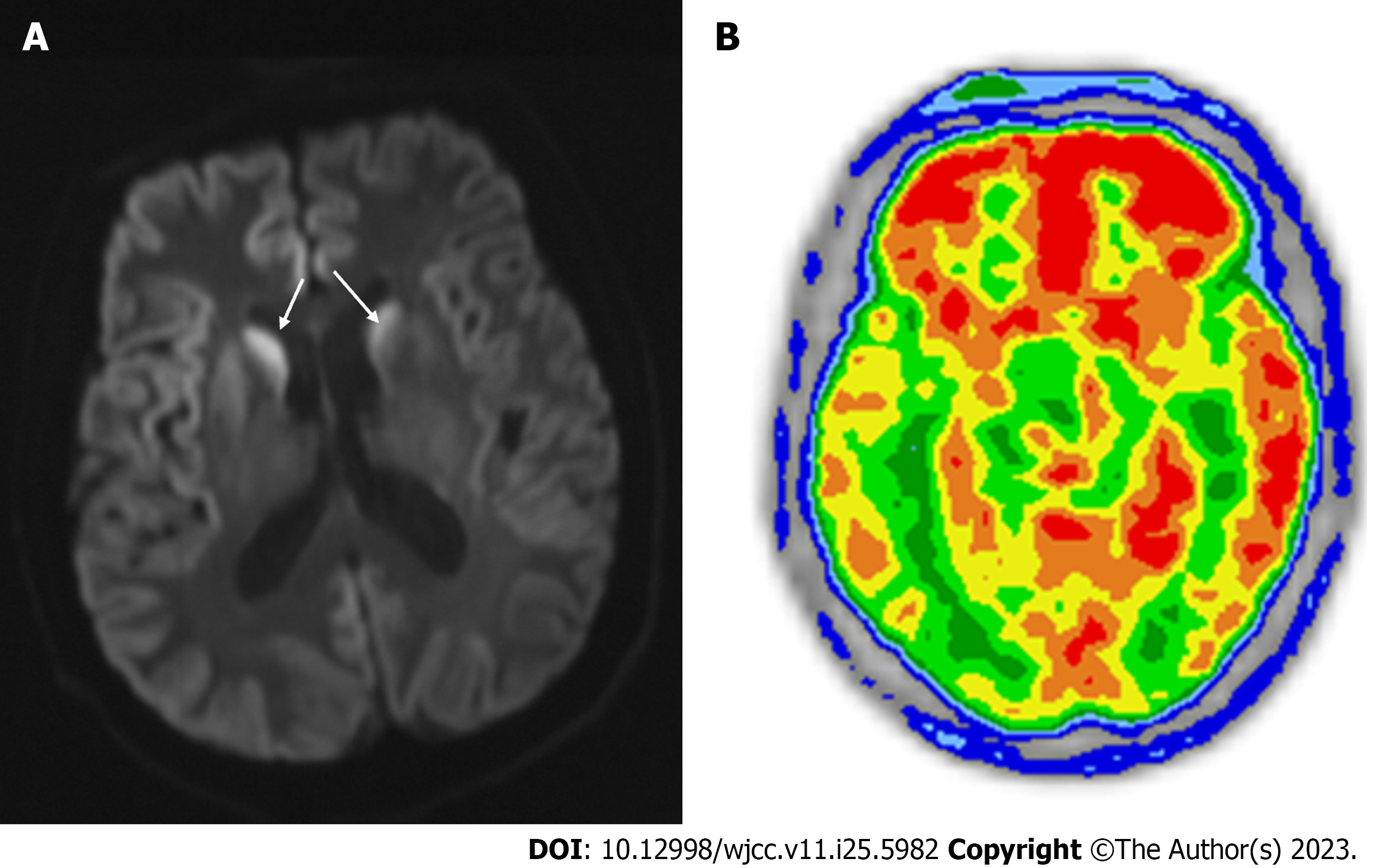
Figure 1 Diffusion-weighted imaging and positron emission tomography/computed tomography of brain.
A: Hyperintense signals observed in bilateral occipital, parietal, temporal, and frontal lobes. Hyperintense signals in bilateral thalamus (white arrow markers); B: Decreased diffuse fluorodeoxyglucose metabolism seen on the right cerebral cortex.
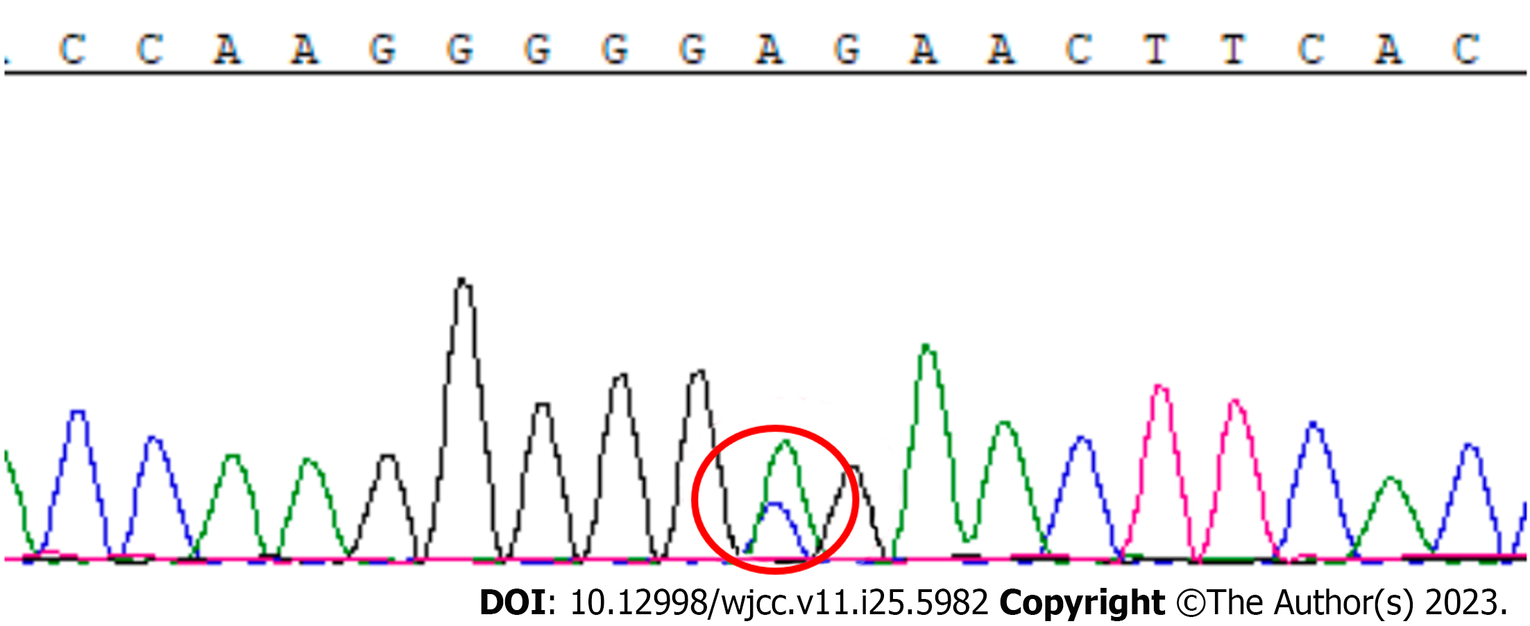
Figure 2 PRNP sequencing outcome.
Red circle marks the presence of a c.A587C base heterozygous change in the second exon of PRNP.
- Citation: Zhang YK, Liu JR, Yin KL, Zong Y, Wang YZ, Cao YM. Creutzfeldt-Jakob disease presenting as Korsakoff syndrome caused by E196A mutation in PRNP gene: A case report. World J Clin Cases 2023; 11(25): 5982-5987
- URL: https://www.wjgnet.com/2307-8960/full/v11/i25/5982.htm
- DOI: https://dx.doi.org/10.12998/wjcc.v11.i25.5982


