Copyright
©The Author(s) 2023.
World J Clin Cases. Sep 6, 2023; 11(25): 5962-5969
Published online Sep 6, 2023. doi: 10.12998/wjcc.v11.i25.5962
Published online Sep 6, 2023. doi: 10.12998/wjcc.v11.i25.5962
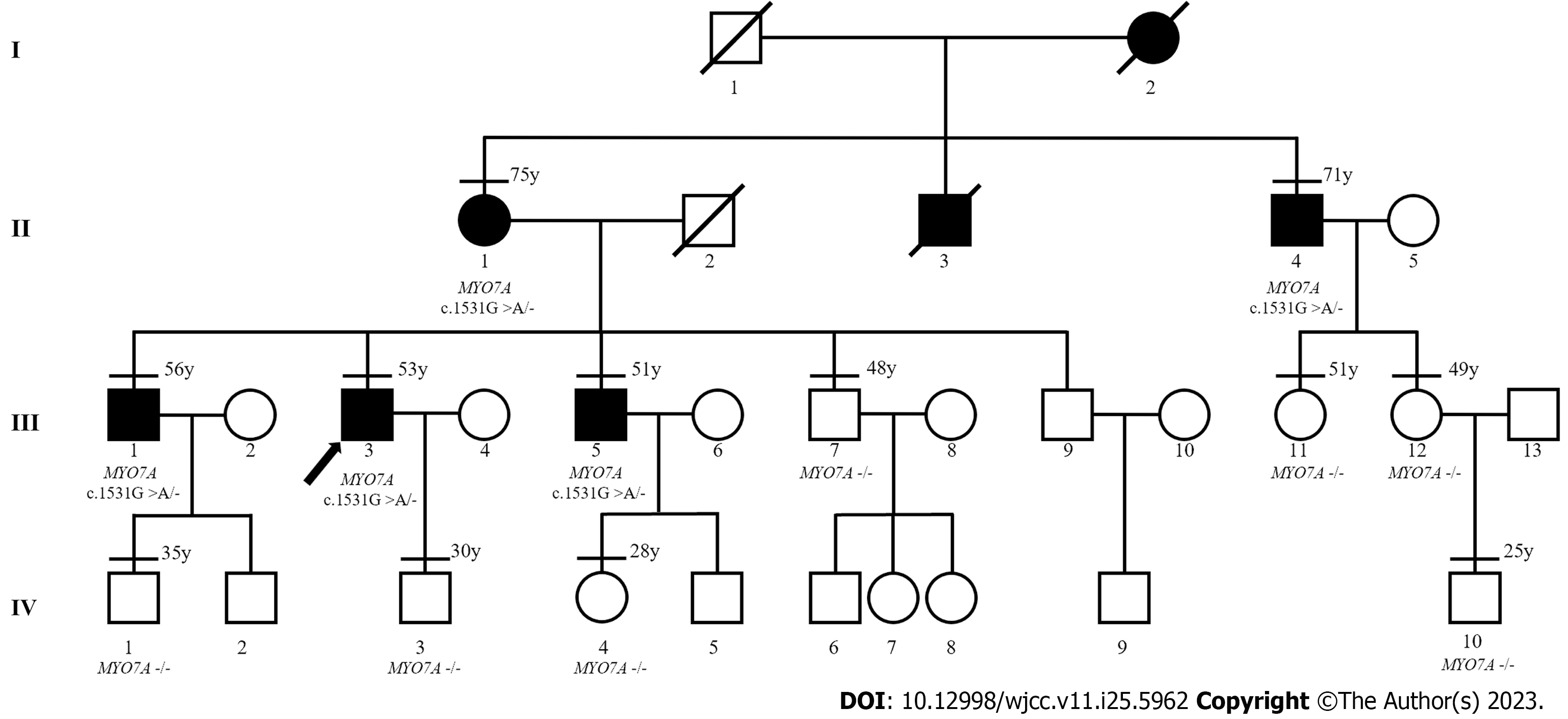
Figure 1 Pedigree of the DFNA11 family.
The arrow indicates the proband. Horizontal lines above the individuals indicate that genetic testing was performed. The age of each subject at the time of genetic testing is listed on the top-right region of each symbol. The genotype of MYO7A for each individual is indicated below the symbol, heterozygous mutant: c.1531G>A/- or wild type: -/-.
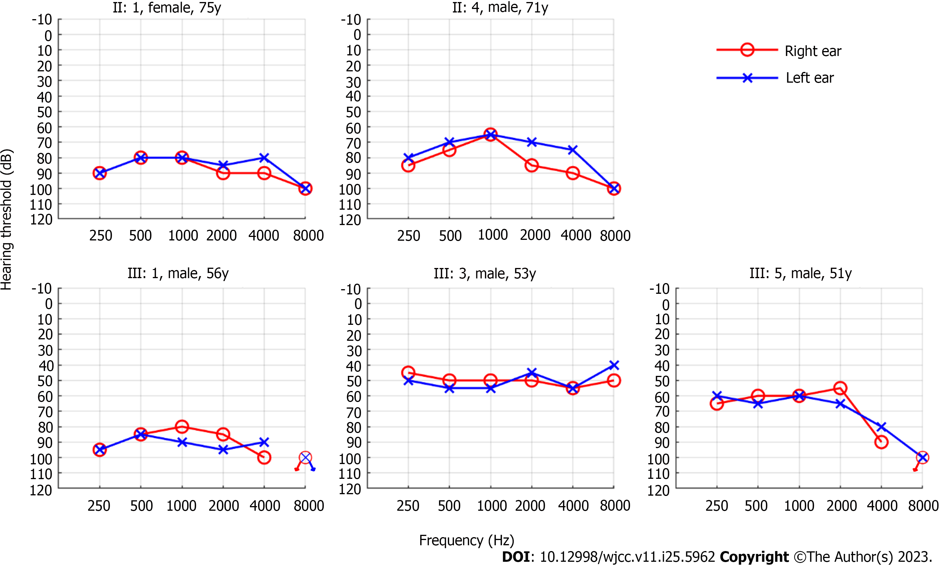
Figure 2 Pure-tone audiogram of the five affected members in this family.
Each person presented bilateral symmetrical moderate-to-profound sensorineural hearing loss, with audiograms that were either flat or slightly downward-sloping at high frequencies. Specifically, III:1 had particularly profound hearing loss owing to occupational noise exposure.
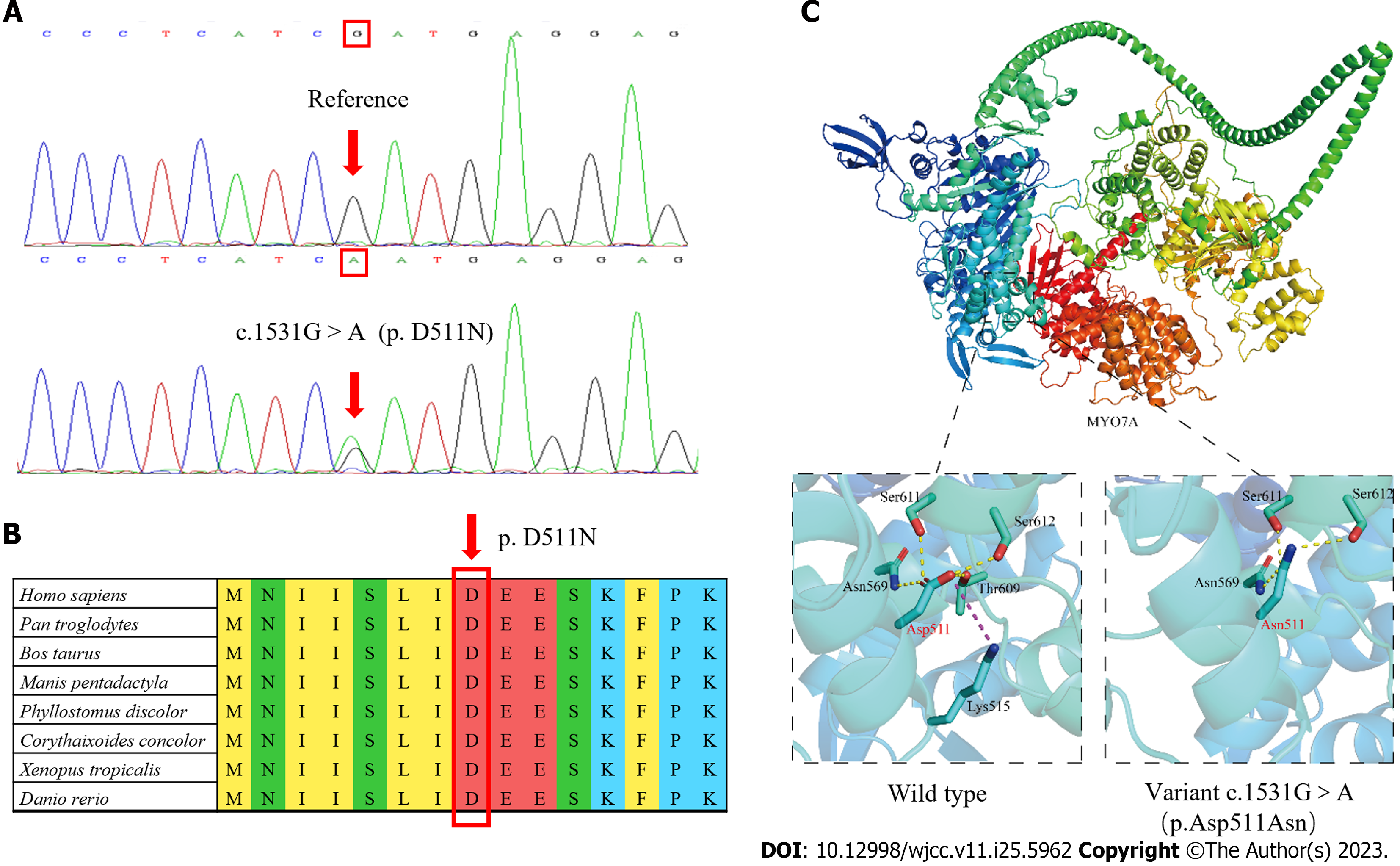
Figure 3 Location of nucleotide changes and functional analysis of the variant.
A: DNA sequence chromatograms. Arrows indicate the site of the mutation, which results in the p.D511N variant; B: Evolutionary conservation of Asp at position 511 (indicated by the arrow) on the MYO7A protein; C: The wild-type and variant (p.D511N) of the MYO7A protein. Yellow dashed lines represent hydrogen bonds between amino acids, and the purple dashed line represent electrostatic interaction between amino acids.
- Citation: Xia CF, Yan R, Su WW, Liu YH. Autosomal dominant non-syndromic hearing loss caused by a novel mutation in MYO7A: A case report and review of the literature. World J Clin Cases 2023; 11(25): 5962-5969
- URL: https://www.wjgnet.com/2307-8960/full/v11/i25/5962.htm
- DOI: https://dx.doi.org/10.12998/wjcc.v11.i25.5962


