Copyright
©The Author(s) 2023.
World J Clin Cases. Sep 6, 2023; 11(25): 5934-5940
Published online Sep 6, 2023. doi: 10.12998/wjcc.v11.i25.5934
Published online Sep 6, 2023. doi: 10.12998/wjcc.v11.i25.5934
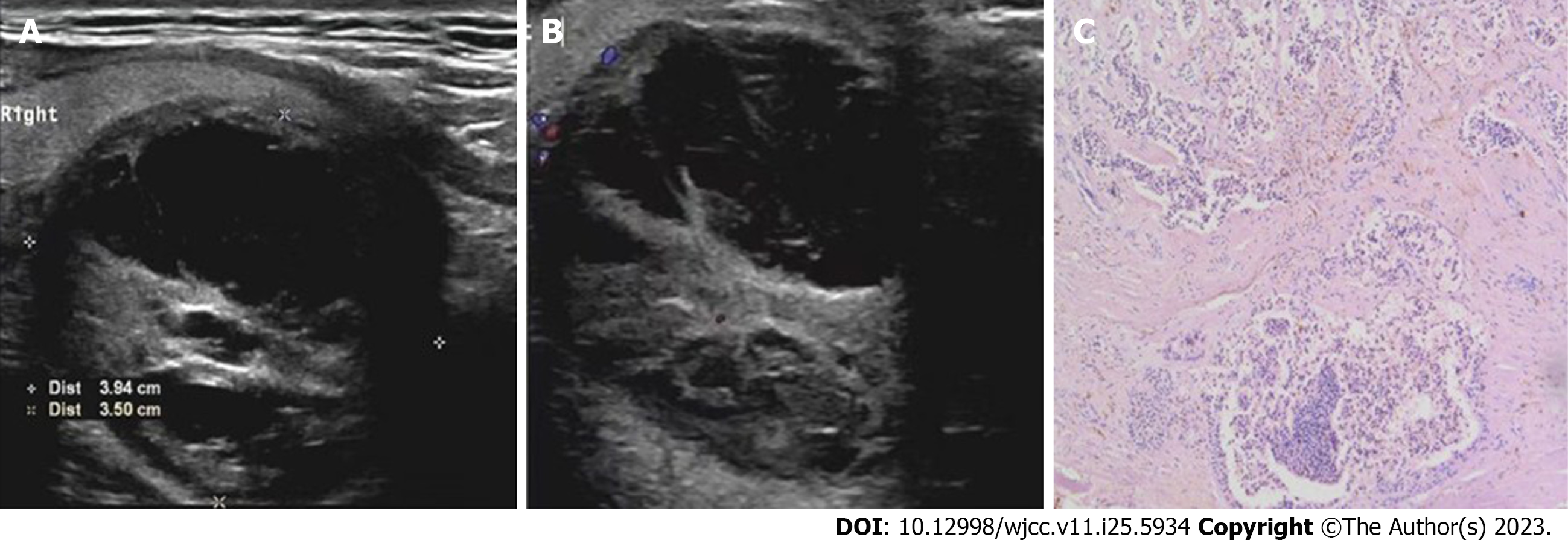
Figure 1 Parathyroid carcinoma of the lower pole of the thyroid on the right side in a 59-year-old man.
A: Doppler ultrasound revealed a complex nodule (with mixed echogenicity), i.e., with solid and fluid components; B: Color doppler flow imaging indicated punctate blood flow signals were detected in the solid part of the nodule and its periphery; C: The neoplastic chief cells of the parathyroid glands were arranged in solid sheets, nests and acini, indicating diffuse infiltrative growth.
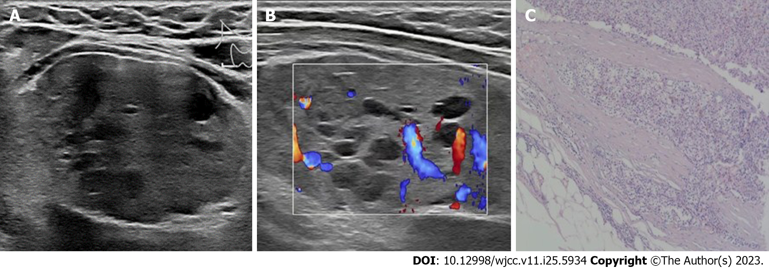
Figure 2 Parathyroid adenocarcinoma of the lower right lobe of the thyroid gland in a 28-year-old woman.
A: Doppler ultrasound revealed a solid nodule with small internal cystic foci; B: Color doppler flow imaging indicated multiple streaks of blood flow signals were observed in the solid part of the nodule; C: Parathyroid chief cells with neoplastic hyperplasia; some of the tumor cells were atypical, and the tumor capsule was of different thicknesses with focal capsule invasion.
Figure 3 Parathyroid adenocarcinoma of the lower right lobe of the thyroid gland in a 65-year-old woman.
A: Doppler ultrasound revealed a solid nodule with an inhomogeneous echostructure; B: Color doppler flow imaging indicated multiple spots and strips of blood flow signals were observed in the nodules; C: Parathyroid tumor with an unclear local capsule. A vascular tumor thrombus can be seen.
- Citation: Shi C, Lu N, Yong YJ, Chu HD, Xia AJ. Parathyroid carcinoma: Three case reports. World J Clin Cases 2023; 11(25): 5934-5940
- URL: https://www.wjgnet.com/2307-8960/full/v11/i25/5934.htm
- DOI: https://dx.doi.org/10.12998/wjcc.v11.i25.5934


