Copyright
©The Author(s) 2023.
World J Clin Cases. Sep 6, 2023; 11(25): 5910-5918
Published online Sep 6, 2023. doi: 10.12998/wjcc.v11.i25.5910
Published online Sep 6, 2023. doi: 10.12998/wjcc.v11.i25.5910
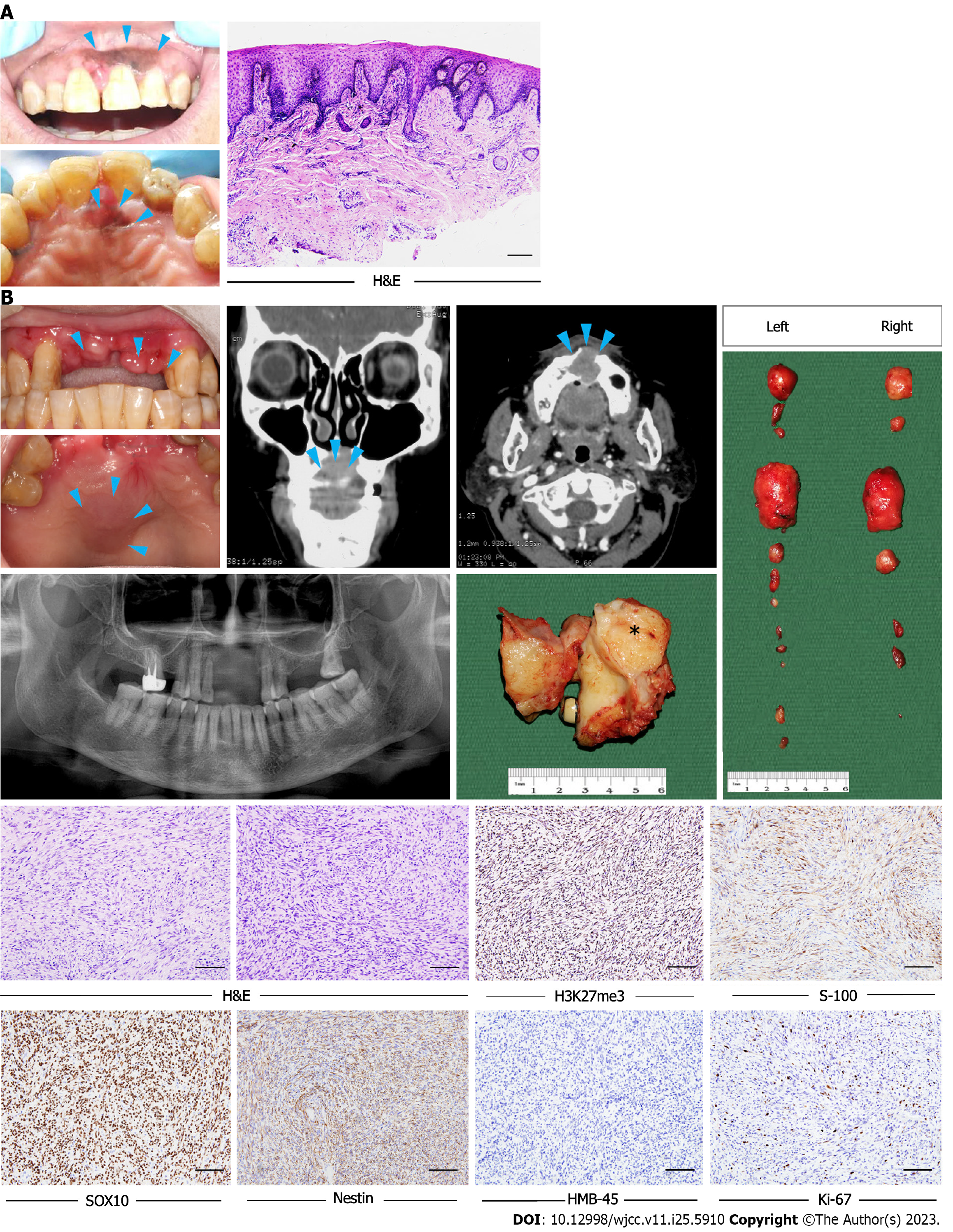
Figure 1 Clinicopathological characteristics of the patient in case 1.
A: Clinical and pathological presentations of the patient during the first visit to our hospital. A macular brown to black lesion was on the anterior maxillary gingiva and incisive foramen region (arrows). Histopathological image revealed the lesion was black macule; B: Clinicopathological presentations of the patient during the second visit to our hospital. A multinodular mass was noted on the anterior maxillary gingiva, and a local swelling was on the anterior to median portion of the palate. Coronal head computed tomography scan shows soft tissue masses in the front of the upper jaw with unclear boundary. The masses involved the post-surgical defect in the incisor region and there was lytic destruction of the underlying bone (arrows). Enlarged lymph nodes and the gross specimen of the lesion after the surgery. H&E characteristics of the case confirmed the diagnosis was malignant peripheral nerve sheath tumor. Tumor cells show partial loss of H3K27me3 and positive stain for S-100, SOX10, nestin, and negative stain for HMB-45. The Ki-67 index is about 10%. Scale bar: 250 μm (A), 50 μm (B).
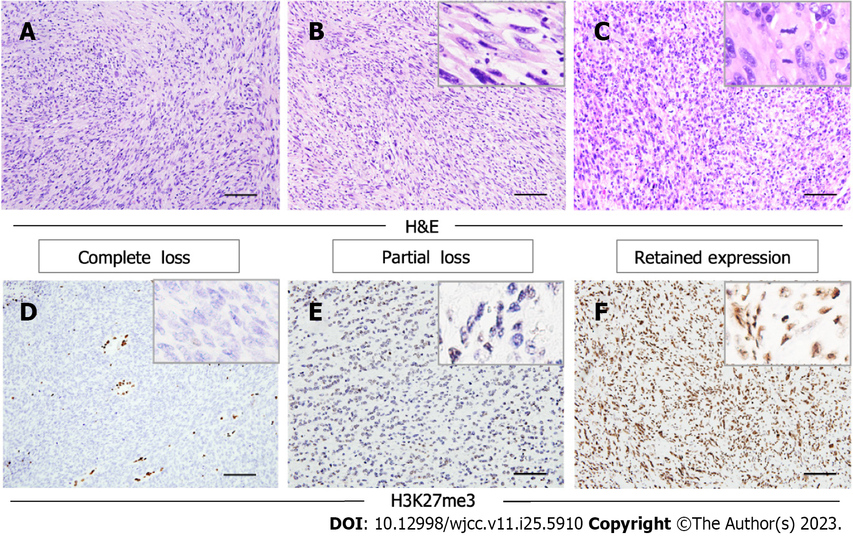
Figure 2 Histological characteristics of malignant peripheral nerve sheath tumor (MPNST) and immunohistochemical analysis of H3K27me3 in MPNST sections.
A: The lesion cells display predominantly spindle in an architectural pattern; B: The cells are enlongated and tends to be hyperchromatic with scant cytoplasm; C: There is severe cytologic atypia and nuclear pleomorphism, with focally brisk mitotic activity; D: Complete loss of H3K27me3 expression on sections. Staining of blood vessels and inflammatory cells is the internal positive controls; E: Partial loss of H3K27me3 expression on the section; F: Strong expression of H3K27me3 is retained in some MPNST cases. Scale bars: 50 μm.
- Citation: Li L, Ma XK, Gao Y, Wang DC, Dong RF, Yan J, Zhang R. Clinicopathological study of malignant peripheral nerve sheath tumors in the head and neck: Case reports and review of literature. World J Clin Cases 2023; 11(25): 5910-5918
- URL: https://www.wjgnet.com/2307-8960/full/v11/i25/5910.htm
- DOI: https://dx.doi.org/10.12998/wjcc.v11.i25.5910


