Copyright
©The Author(s) 2023.
World J Clin Cases. Aug 26, 2023; 11(24): 5804-5810
Published online Aug 26, 2023. doi: 10.12998/wjcc.v11.i24.5804
Published online Aug 26, 2023. doi: 10.12998/wjcc.v11.i24.5804
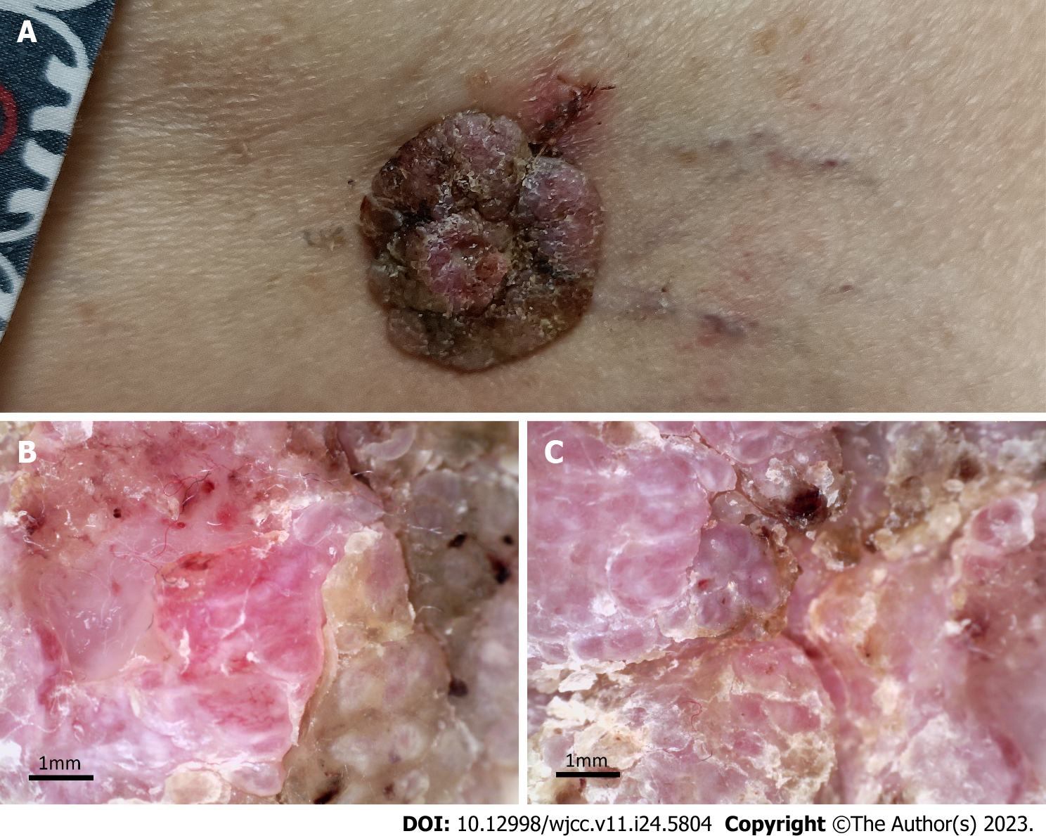
Figure 1 Clinical image and dermoscopic examination of the tumour.
A: Clinical picture; B and C: Dermoscopic images of the lesion. (B) ×50, (C) ×50.
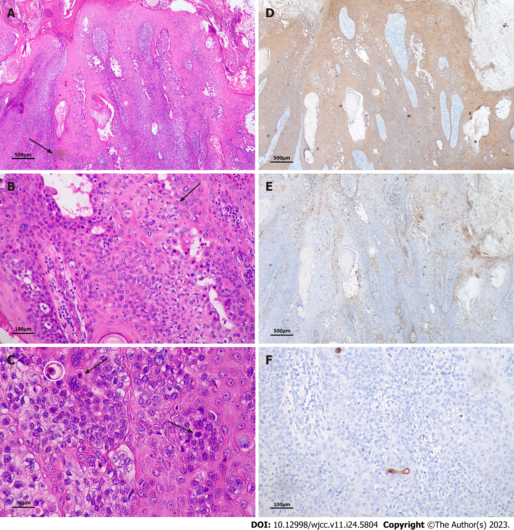
Figure 2 Histopathological and immunohistochemical analysis.
A-C: Haematoxylin and eosin staining of resected specimen (A) ×40, (B) ×200, (C) ×400. A: Blunted epithelial feet (white arrow); B: Neoplastic cells were less stained than the surrounding epithelium (black arrow); C: Dyskeratotic cells (white circle) and mitotic figures (black arrows); D: Immunohistochemical staining showed positivity for CK5/6 (×40); E: EMA (×40); F: CEA was only expressed in ductal structures ×200.
- Citation: Yang YF, Wang R, Xu H, Long WG, Zhao XH, Li YM. Malignant form of hidroacanthoma simplex: A case report. World J Clin Cases 2023; 11(24): 5804-5810
- URL: https://www.wjgnet.com/2307-8960/full/v11/i24/5804.htm
- DOI: https://dx.doi.org/10.12998/wjcc.v11.i24.5804


