Copyright
©The Author(s) 2023.
World J Clin Cases. Aug 26, 2023; 11(24): 5789-5796
Published online Aug 26, 2023. doi: 10.12998/wjcc.v11.i24.5789
Published online Aug 26, 2023. doi: 10.12998/wjcc.v11.i24.5789
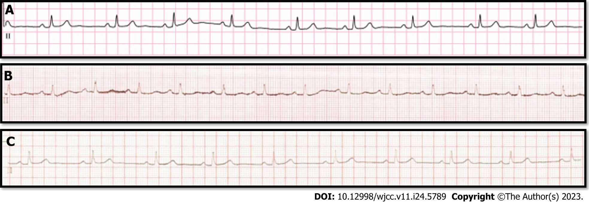
Figure 1 Perioperative electrocardiogram of the patient.
A: Preoperative electrocardiogram (ECG), sinus rhythm, heart rate (HR) 57 beats/min; B: Postoperative ECG (postoperative day 0), accelerated junctional rhythm, HR 83 beats/min; C: Postoperative ECG (postoperative day 2), sinus rhythm, HR 58 beats/min.
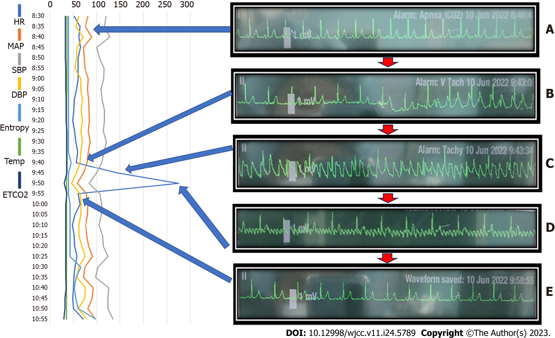
Figure 2 Hemodynamic data and events with drug administration and electrocardiogram in chronological order.
A: Electrocardiogram (ECG) revealed a normal sinus rhythm at the start of the operation; B: First, paroxysmal supraventricular tachycardia pattern arrythmia occurred suddenly during approach to the cervical spine; C: Flutter-like pattern on ECG occurred continuously; D: Adenosine was administered, and the ECG pattern changed to atrial flutter (AF). Diltiazem injection for management of AF; E: Disappearance of flutter-like ECG. MAP: Mean arterial pressure; HR: Heart rate; SBP: Systolic blood pressure; DBP: Diastolic blood pressure; Entropy: Sedation scale; Temp: Body temperature; ETCO2: End-tidal carbon dioxide.
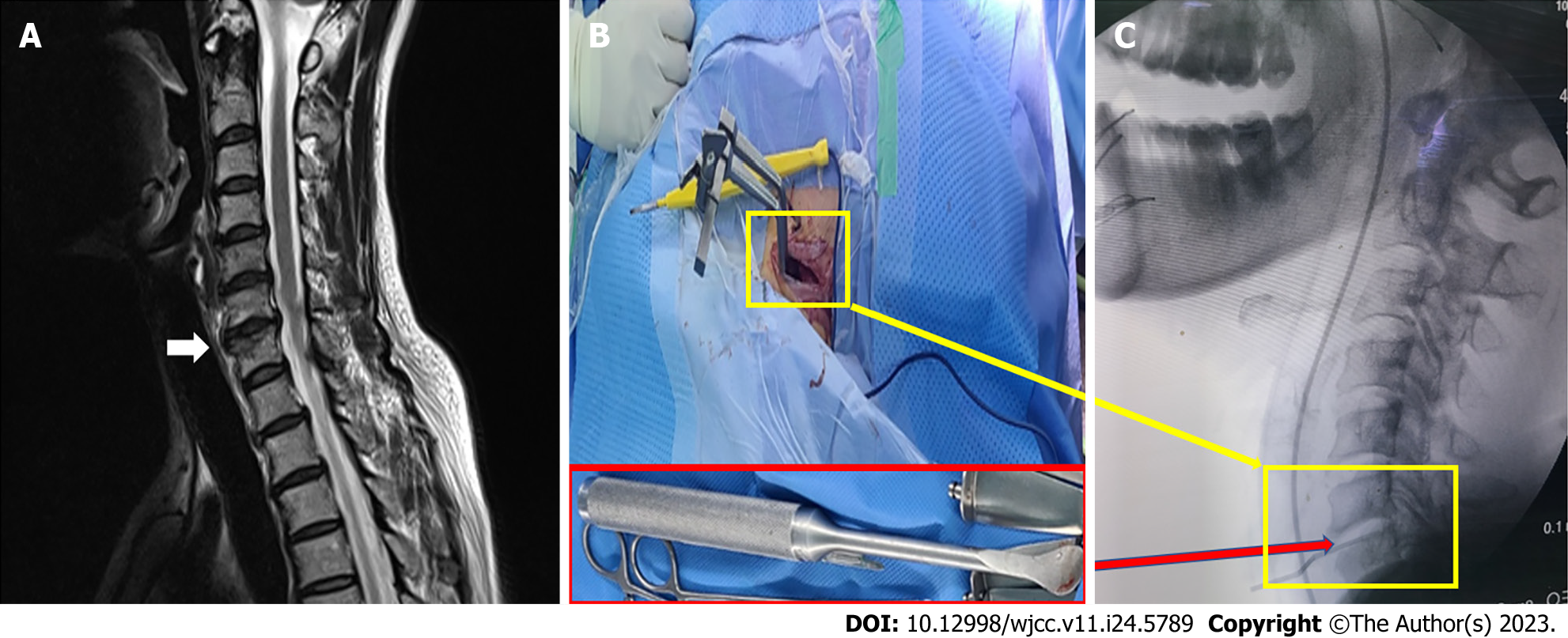
Figure 3 Images of the patient.
A: Magnetic resonance imaging image: C6-7 disc bulging with marginal spur and neural foraminal stenosis of C6-7; B and C: Intraoperative picture with C-arm image: tracing with cervical distractor to expose the surgical site and spinal fusion retraction device used in this operation. Indication device for exposure of C6-7 intervertebral disc space.
- Citation: Seo JH, Cho SY, Park JH, Seo JY, Lee HY, Kim DJ. Intraoperative sudden arrhythmias in cervical spine surgery adjacent to the stellate ganglion: A case report. World J Clin Cases 2023; 11(24): 5789-5796
- URL: https://www.wjgnet.com/2307-8960/full/v11/i24/5789.htm
- DOI: https://dx.doi.org/10.12998/wjcc.v11.i24.5789


