Copyright
©The Author(s) 2023.
World J Clin Cases. Aug 26, 2023; 11(24): 5780-5788
Published online Aug 26, 2023. doi: 10.12998/wjcc.v11.i24.5780
Published online Aug 26, 2023. doi: 10.12998/wjcc.v11.i24.5780
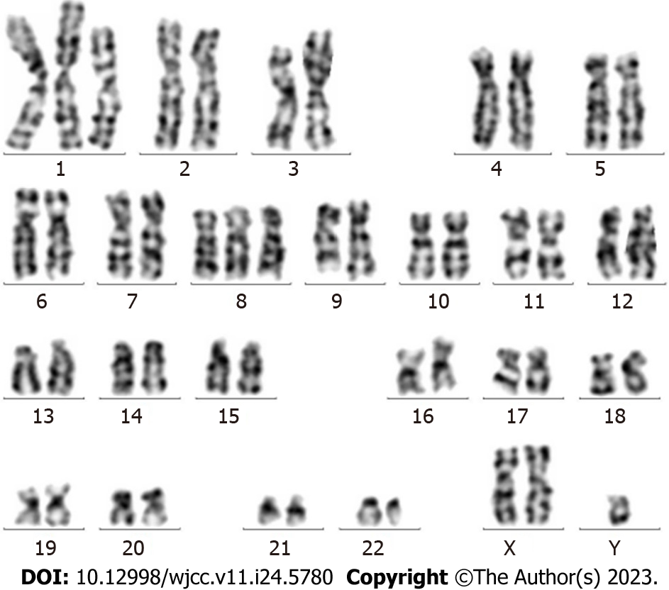
Figure 1 Karyotype analysis of the chronic myelogenous leukemia patient on August 21, 2020.
The patient’s karyotype has transitioned from normal to complex, including the following abnormal karyotypes: 47XY,+X,t(9;22)(q34;q11.2)[6]/49,idem,+1,der(1;7)(q10;q10),+7,+8[2]/49,idem,+1,der(1;7),+8,+10[2]).
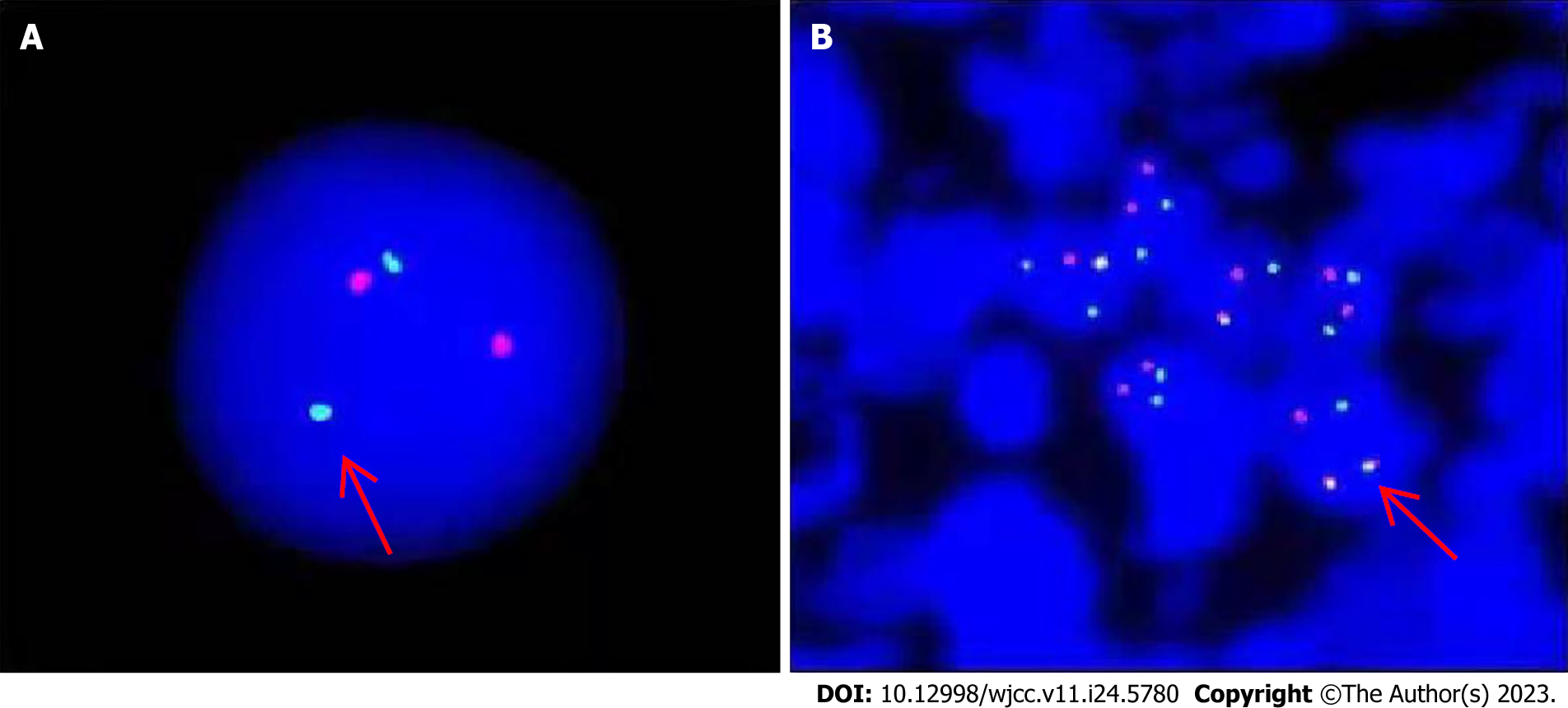
Figure 2 Fluorescence in situ hybridization test of lymph node specimens showed positive BCR-ABL fusion gene in tumor cells.
A: Positive result of single cell BCR-ABL detection; B: Positive result of multicellular BCR-ABL detection.
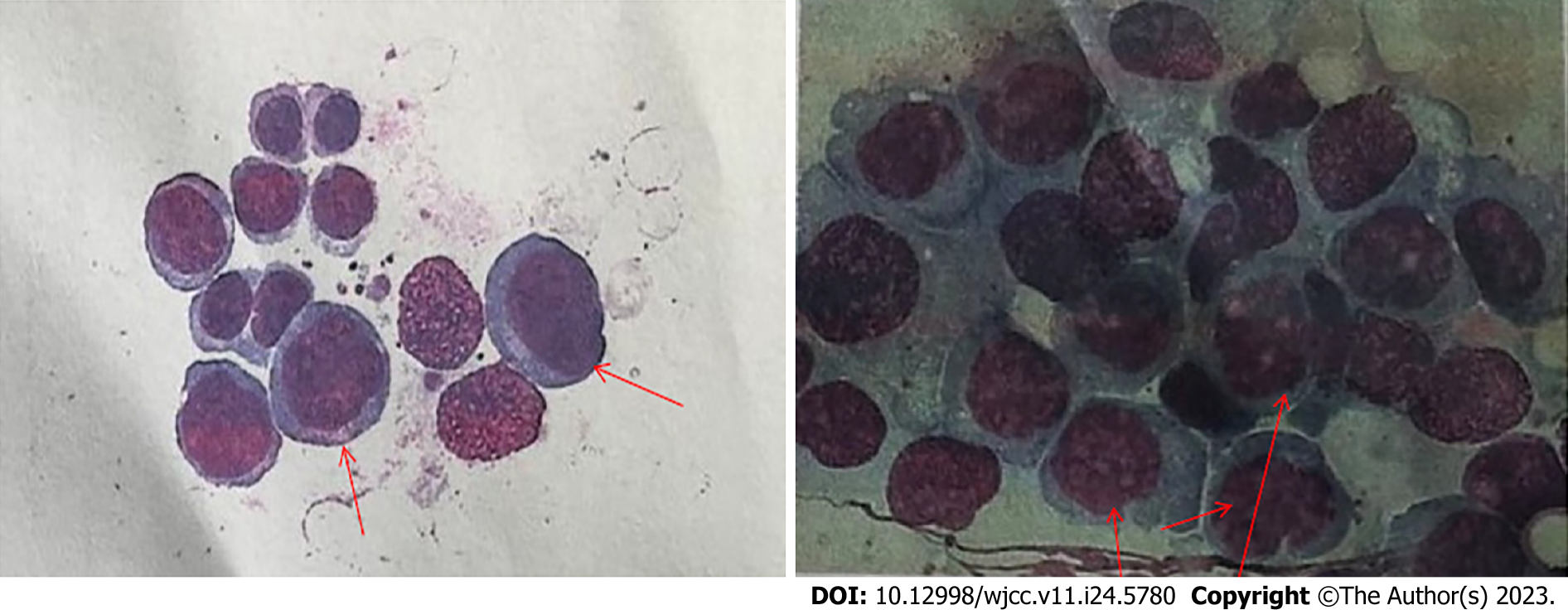
Figure 3 Myelocytic morphology in the patient with erythroid leukemia developing from chronic myelogenous leukemia.
Arrows indicate primitive red blood cells.
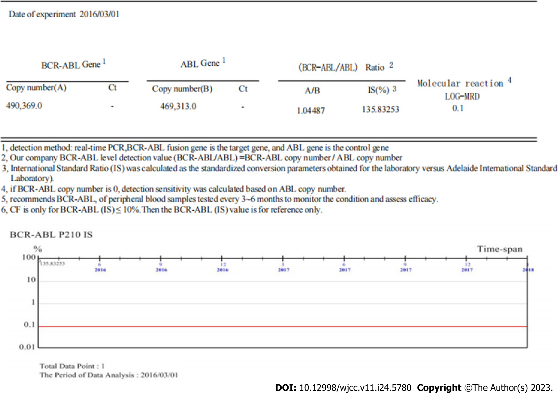
Figure 4 Positive results of BCR-ABLI (p210) fusion gene.
BCR-ABL fusion gene copy number and ratio to internal reference ABL were detected by quantitative real-time polymerase chain reaction.
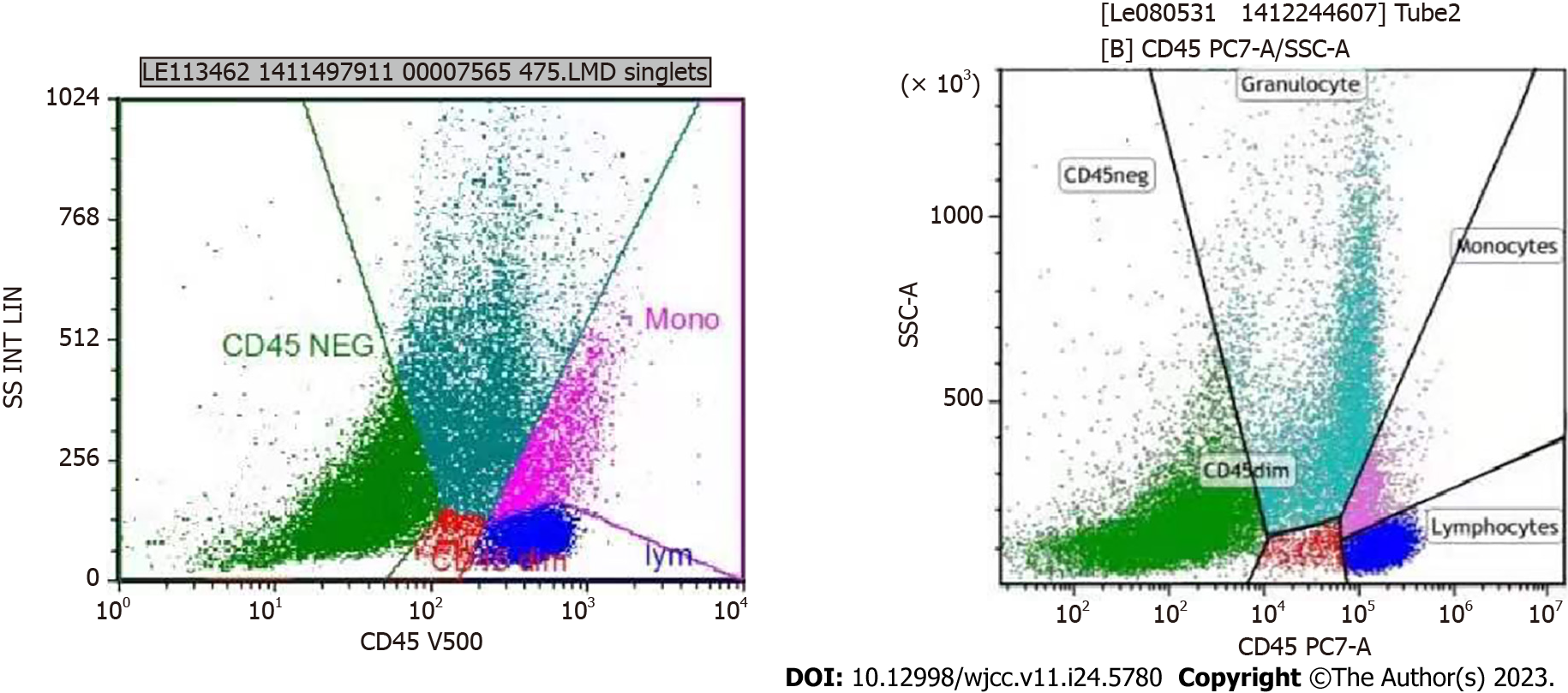
Figure 5 Flow cytometry immunofluorescence analysis of the patient with erythroid leukemia developing from chronic myelogenous leukemia.
The expression characteristics of primitive red blood cell CD antigens in bone marrow progenitor cells were detected by flow cytometry.
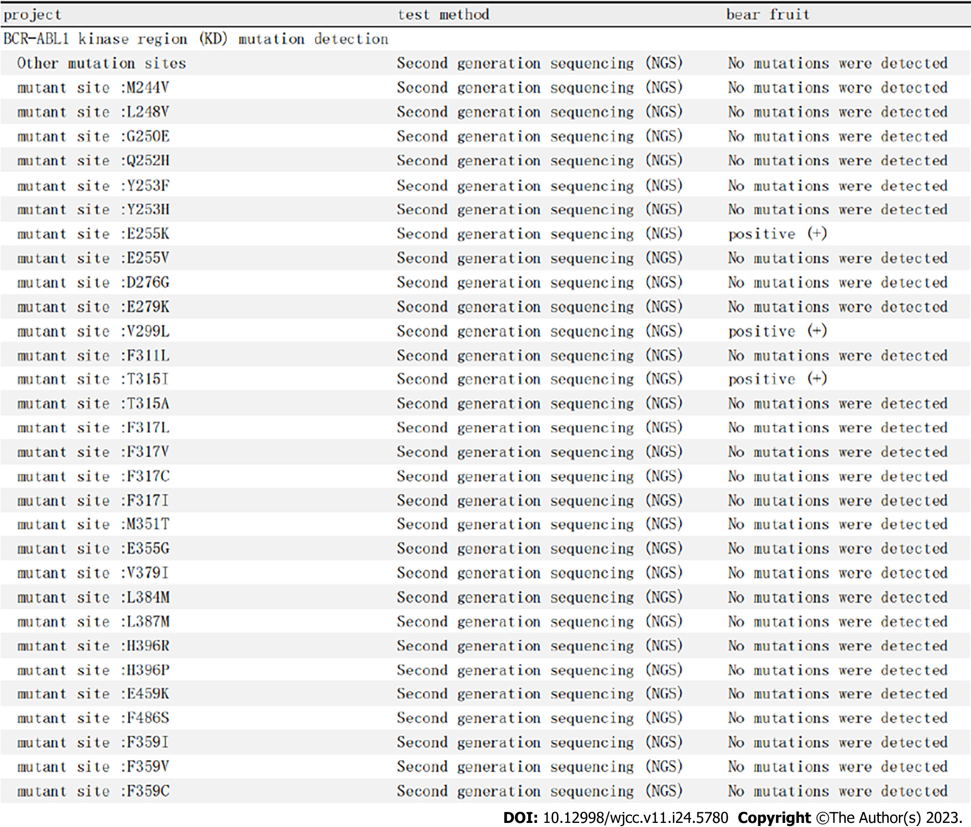
Figure 6 Examination of kinase domain mutations on October 15, 2020.
ABL1 kinase mutation sequence diagram of the patient with erythroid leukemia developing from chronic myelogenous leukemia is shown. Arrows indicate that there are multiple gene mutations in the BCR-ABL kinase region, such as the typical mutation T351I.
- Citation: Wang W, Chen YL, Gou PP, Wu PL, Shan KS, Zhang DL. Focal lymphoblastic transformation of chronic myelogenous leukemia develops into erythroid leukemia: A case report. World J Clin Cases 2023; 11(24): 5780-5788
- URL: https://www.wjgnet.com/2307-8960/full/v11/i24/5780.htm
- DOI: https://dx.doi.org/10.12998/wjcc.v11.i24.5780


