Copyright
©The Author(s) 2023.
World J Clin Cases. Aug 26, 2023; 11(24): 5729-5735
Published online Aug 26, 2023. doi: 10.12998/wjcc.v11.i24.5729
Published online Aug 26, 2023. doi: 10.12998/wjcc.v11.i24.5729
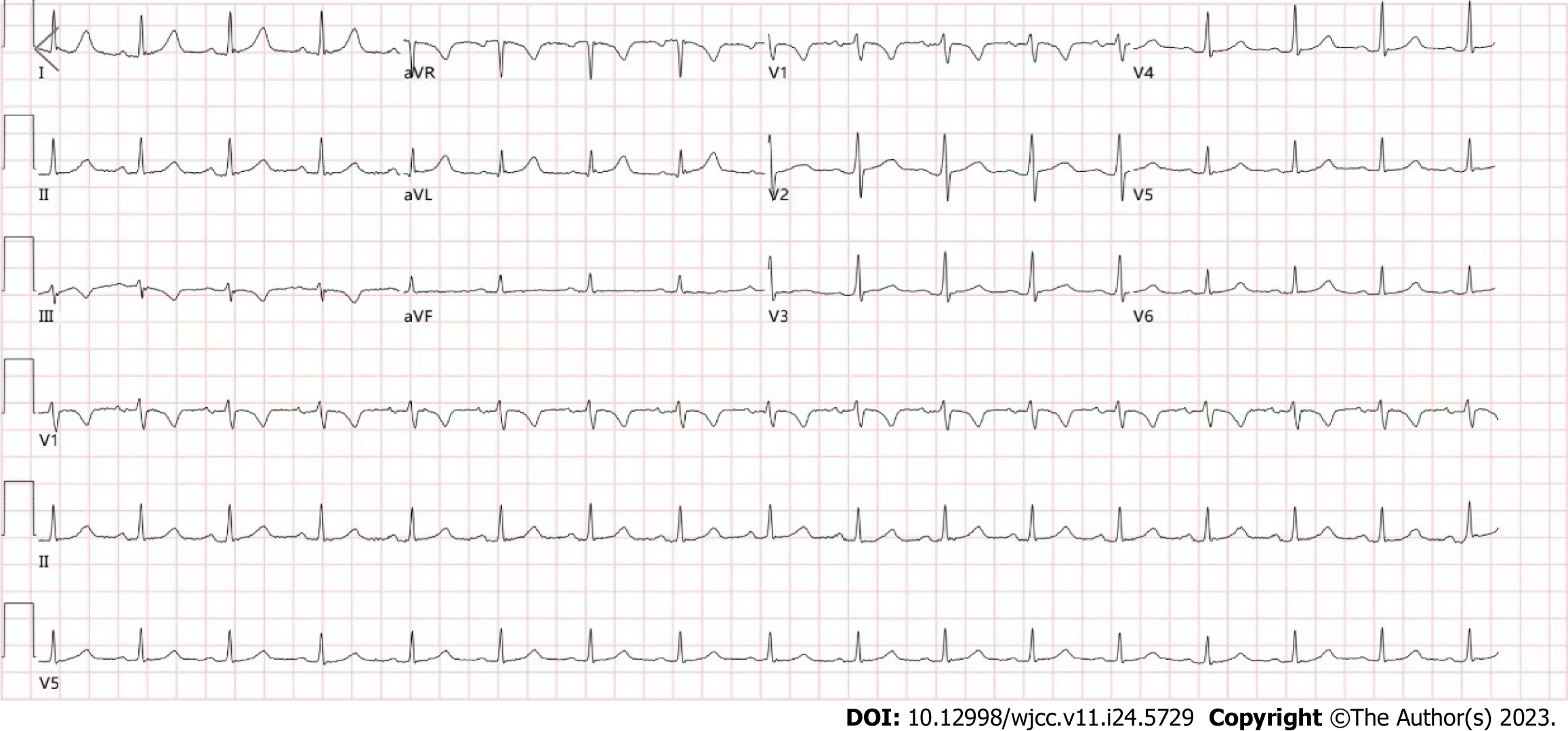
Figure 1 Electrocardiogram findings.
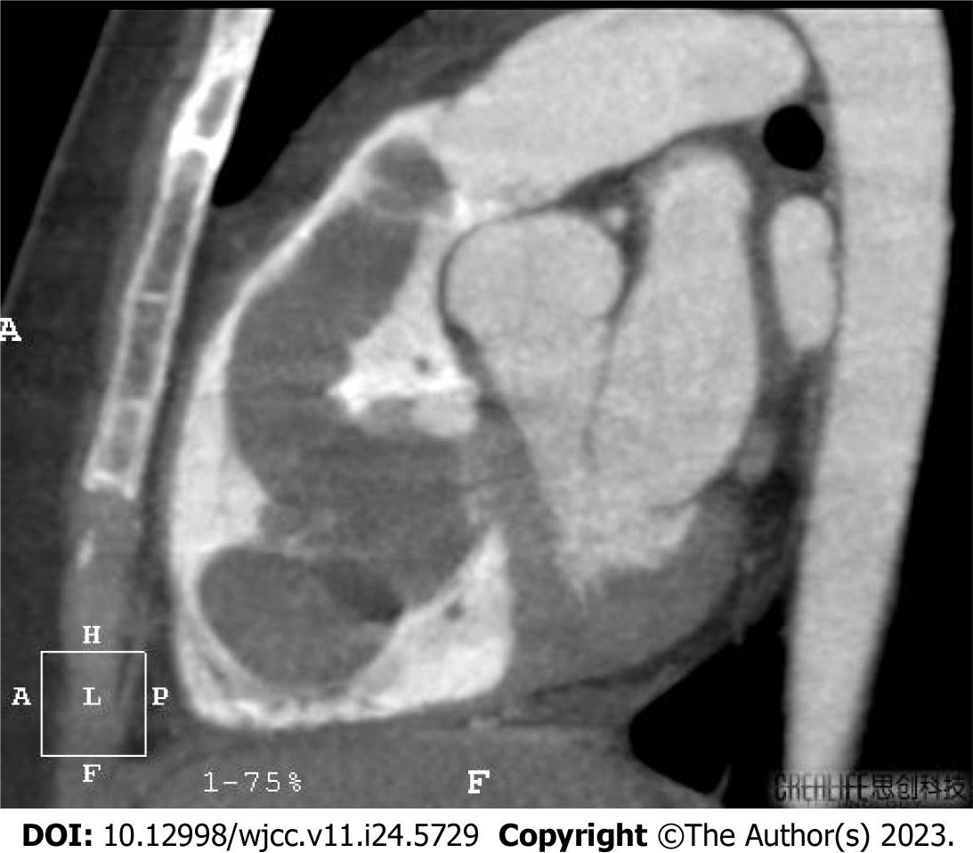
Figure 2 Computed tomography imaging findings.
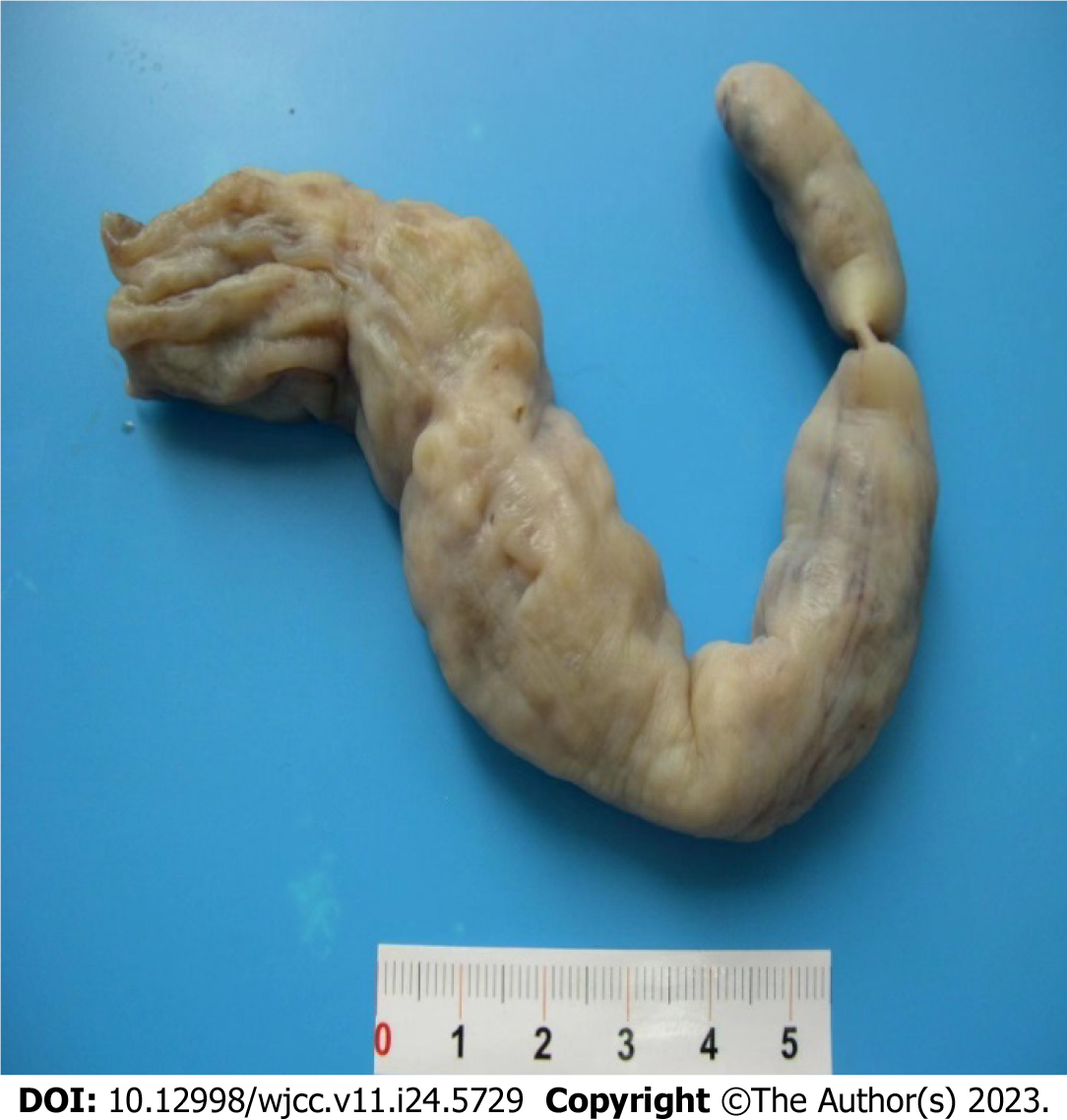
Figure 3 Resected mass.
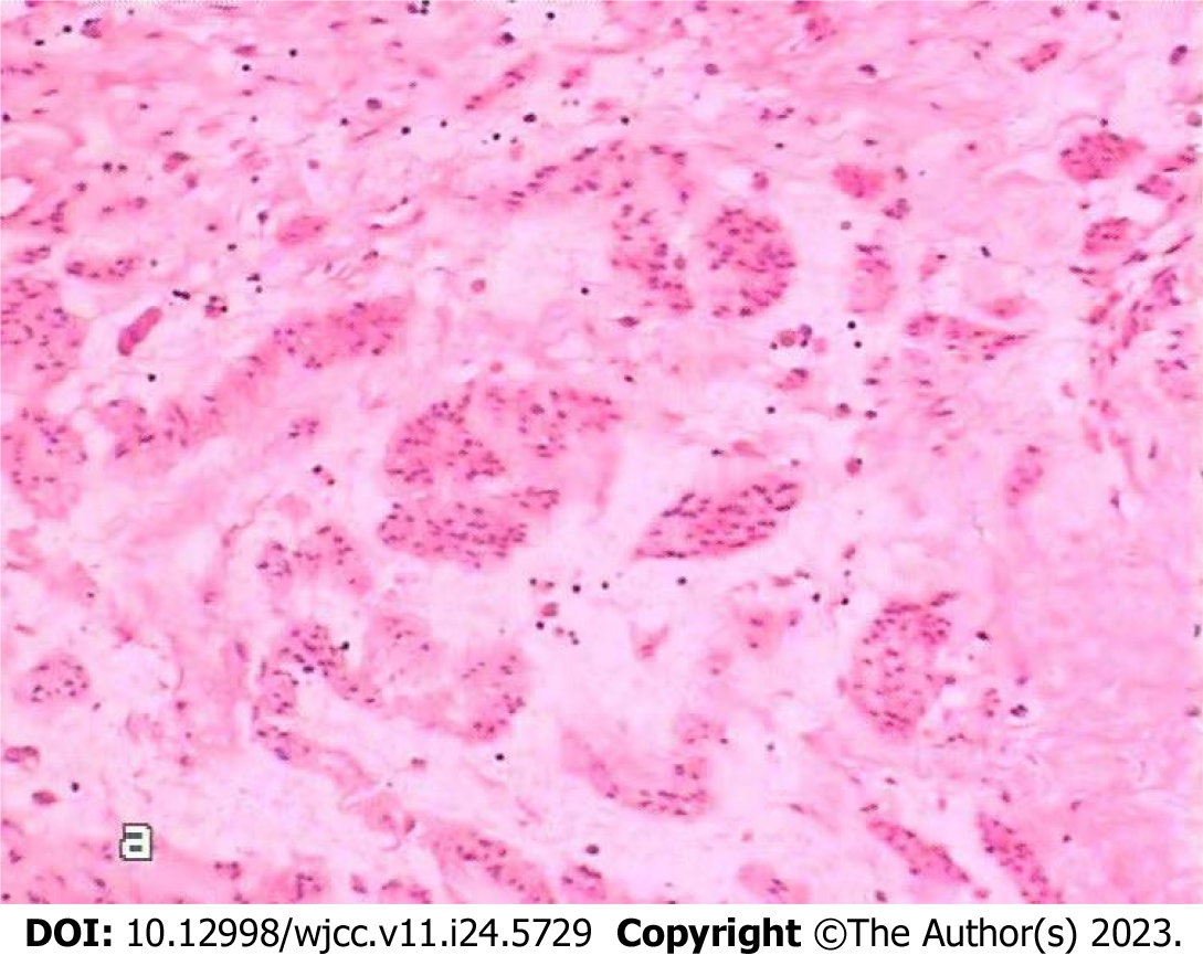
Figure 4 Intravenous leiomyomatosis under a general optical microscope (HE × 10).
- Citation: Huang YQ, Wang Q, Xiang DD, Gan Q. Intravenous leiomyoma of the uterus extending to the pulmonary artery: A case report. World J Clin Cases 2023; 11(24): 5729-5735
- URL: https://www.wjgnet.com/2307-8960/full/v11/i24/5729.htm
- DOI: https://dx.doi.org/10.12998/wjcc.v11.i24.5729


