Copyright
©The Author(s) 2023.
World J Clin Cases. Aug 16, 2023; 11(23): 5615-5621
Published online Aug 16, 2023. doi: 10.12998/wjcc.v11.i23.5615
Published online Aug 16, 2023. doi: 10.12998/wjcc.v11.i23.5615
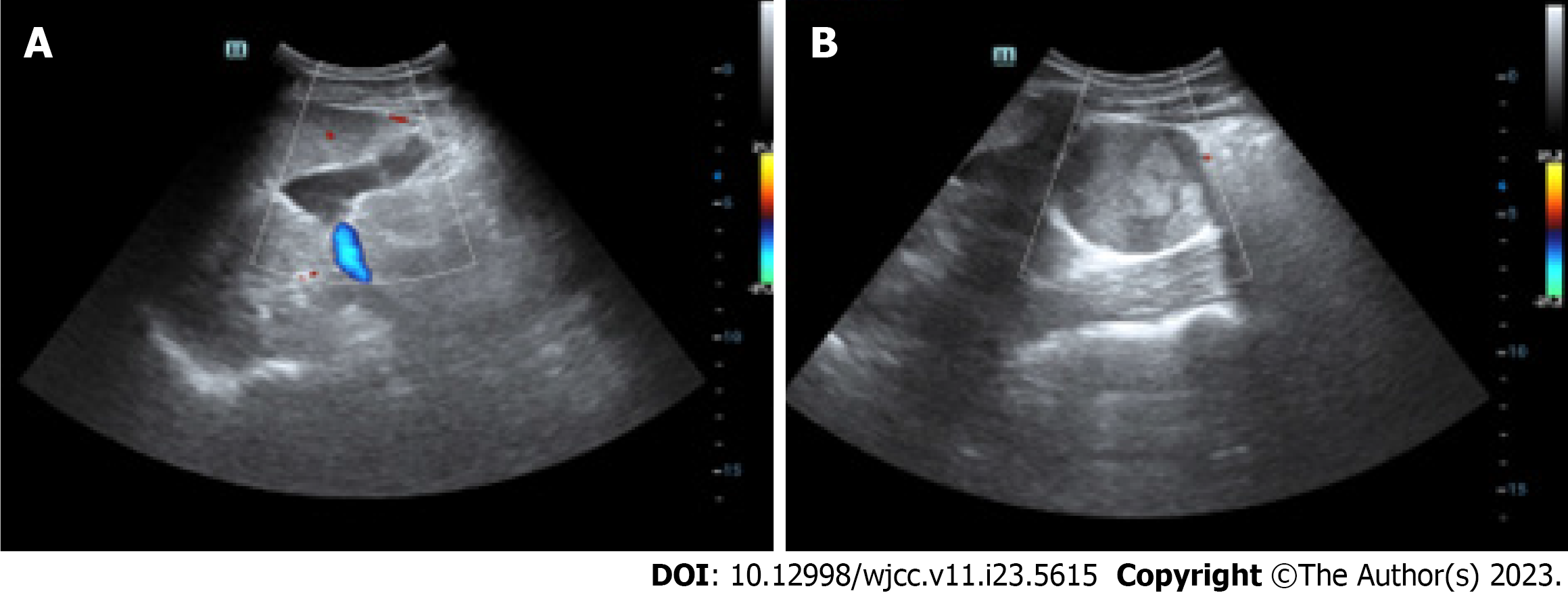
Figure 1 An 18-year-old male with a huge abdominal cyst lesion was diagnosed by trans-abdominal ultrasound.
A: Fluid around the tumor; B: The tumor identified by color Doppler.
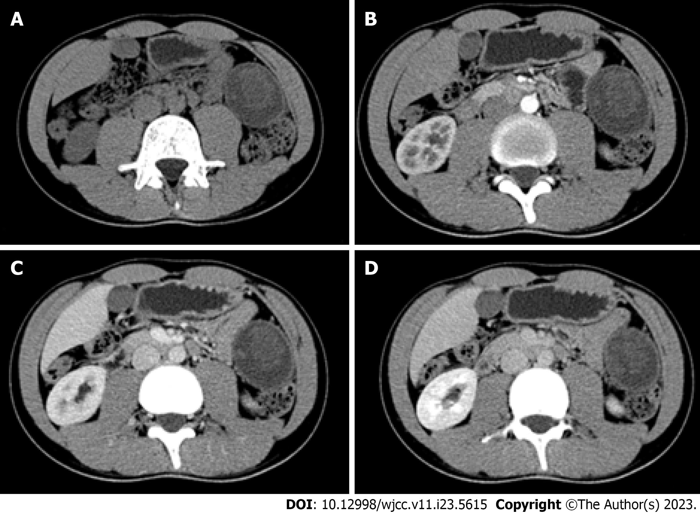
Figure 2 Computed tomography scan images.
A: Plain scan; B: Arterial phase; C: Portal phase; D: Equilibrium phase.
Figure 3 Magnetic resonance imaging scan images.
A: T1 weighted image (T1WI) sequence; B: Coronal position of T2WI sequence; C: T2WI sequence; D: Venous phase of contrast-enhanced magnetic resonance imaging.
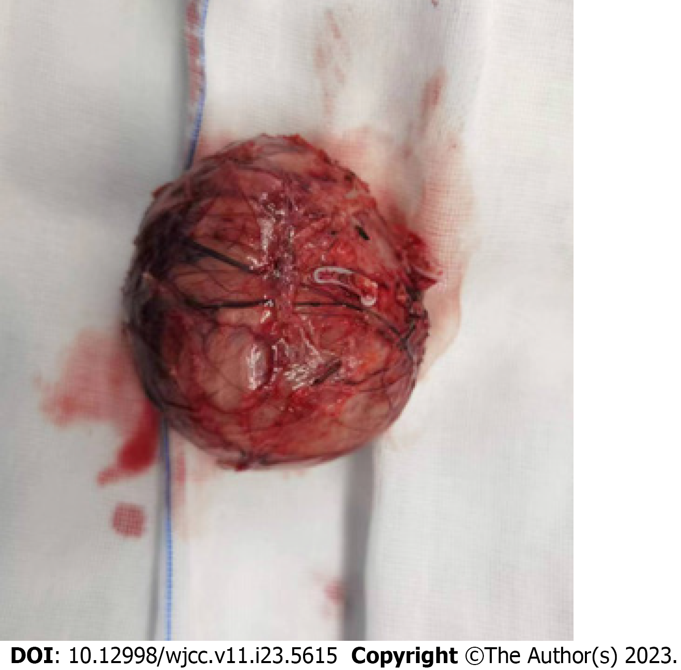
Figure 4 Gross pathology shows the tumor is sac-like, soft in texture, and located at the tail of the pancreas.
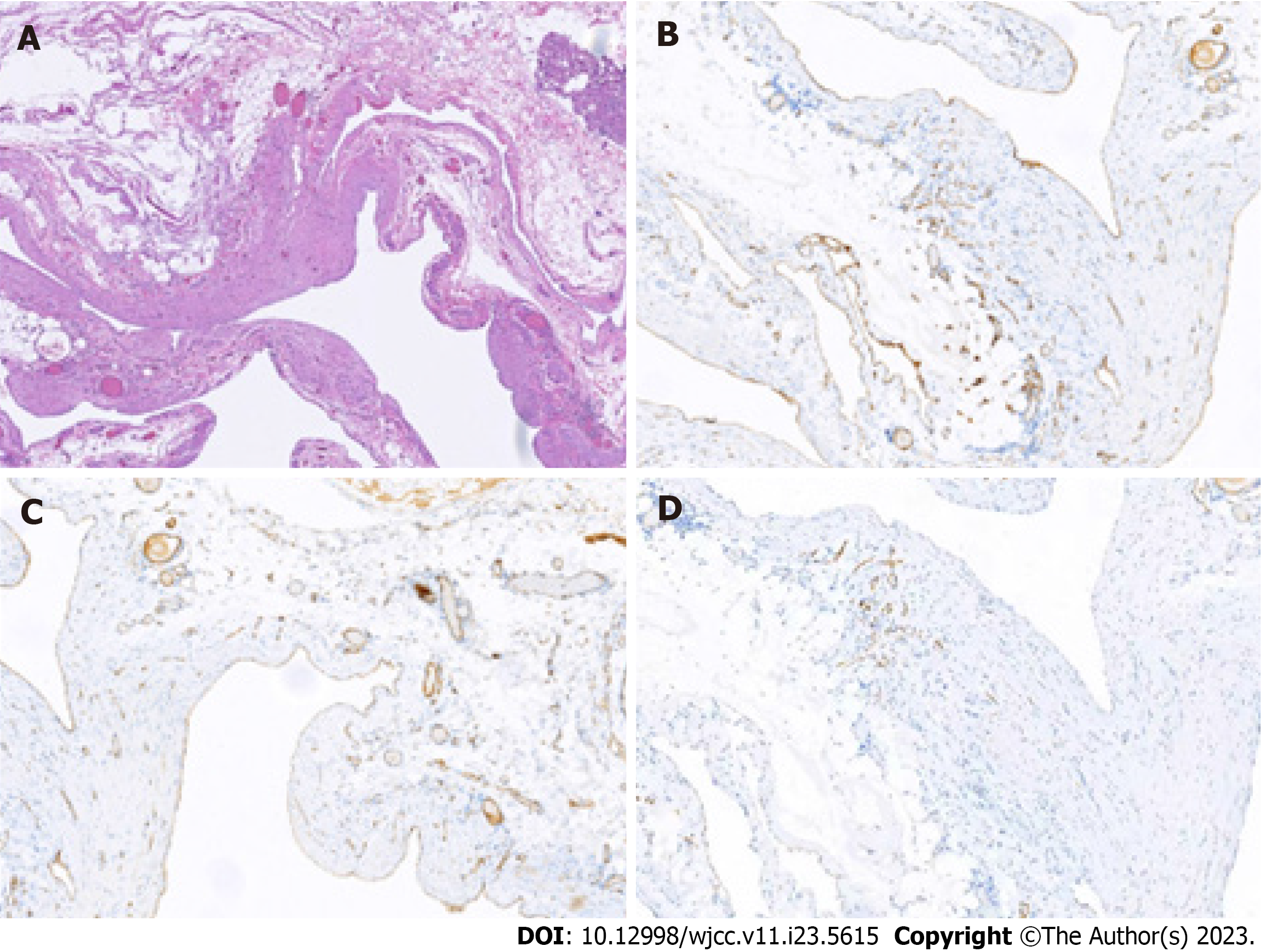
Figure 5 Hematoxylin-eosin staining and immunohistochemical staining of the tumor.
A: Hematoxylin-eosin (HE) staining of the tumor showed proliferative vessels in the fibrous adipose tissue, with different lumen sizes and thicknesses (HE 10); B and C: Immunohistochemical staining showed positive for CD 31 and CD 34; D: D2-40 showed positive on interstitial lymphatic vessels and negative on vascular epithelial cells.
- Citation: Li T. Pancreatic cavernous hemangioma complicated with chronic intracapsular spontaneous hemorrhage: A case report and review of literature. World J Clin Cases 2023; 11(23): 5615-5621
- URL: https://www.wjgnet.com/2307-8960/full/v11/i23/5615.htm
- DOI: https://dx.doi.org/10.12998/wjcc.v11.i23.5615


