Copyright
©The Author(s) 2023.
World J Clin Cases. Aug 16, 2023; 11(23): 5525-5529
Published online Aug 16, 2023. doi: 10.12998/wjcc.v11.i23.5525
Published online Aug 16, 2023. doi: 10.12998/wjcc.v11.i23.5525
Figure 1 Computed tomography.
An emphysematous sloughed tissue within the urinary bladder was observed. A: Front view; B: Side view.
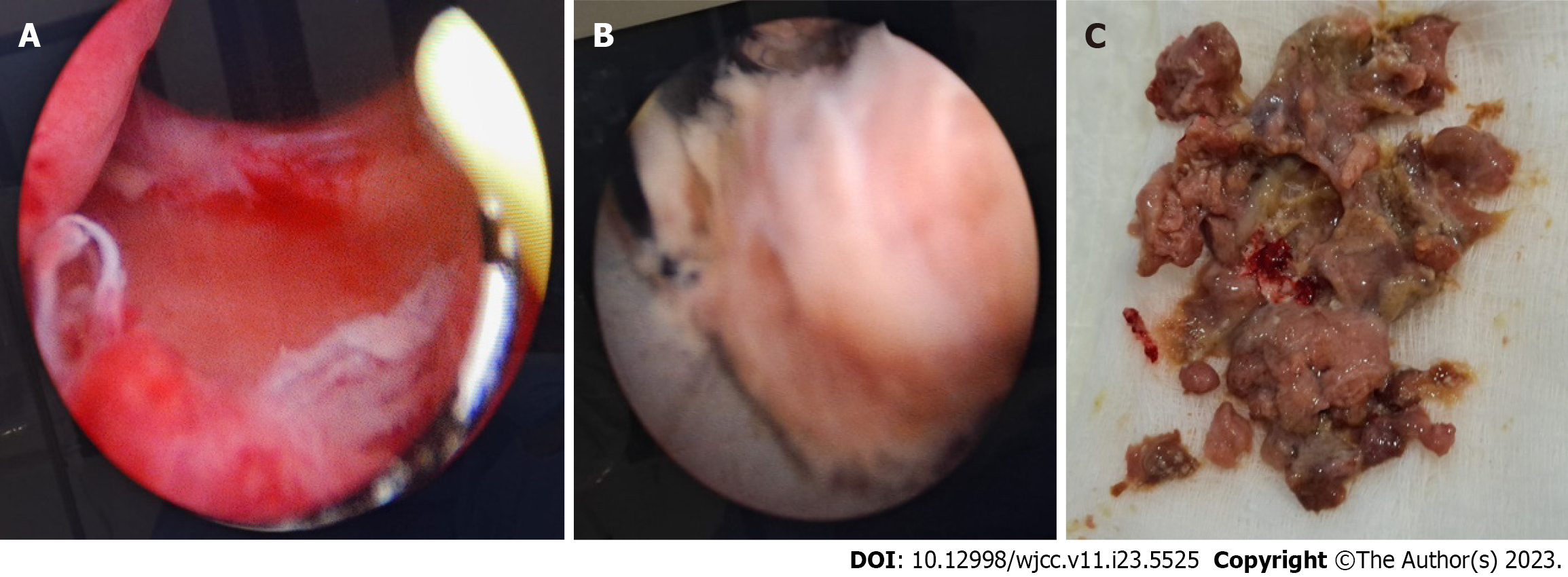
Figure 2 Transurethral removal.
A: Cystoscopy revealed a wide, unobstructed prostatic urethra; B: Cystoscopic view of sloughed Prostatic tissue post Rezum therapy, removed with the loop of a resectoscope; C: Postoperative view of the sloughed Prostatic tissue.
Figure 3
Uroflowmetry after removal of sloughed prostatic tissue.
- Citation: Alnazari M, Bakhsh A, Rajih ES. Emphysematous sloughed floating ball after prostate water vaporization Rezum: A case report. World J Clin Cases 2023; 11(23): 5525-5529
- URL: https://www.wjgnet.com/2307-8960/full/v11/i23/5525.htm
- DOI: https://dx.doi.org/10.12998/wjcc.v11.i23.5525


