Copyright
©The Author(s) 2023.
World J Clin Cases. Aug 6, 2023; 11(22): 5373-5381
Published online Aug 6, 2023. doi: 10.12998/wjcc.v11.i22.5373
Published online Aug 6, 2023. doi: 10.12998/wjcc.v11.i22.5373
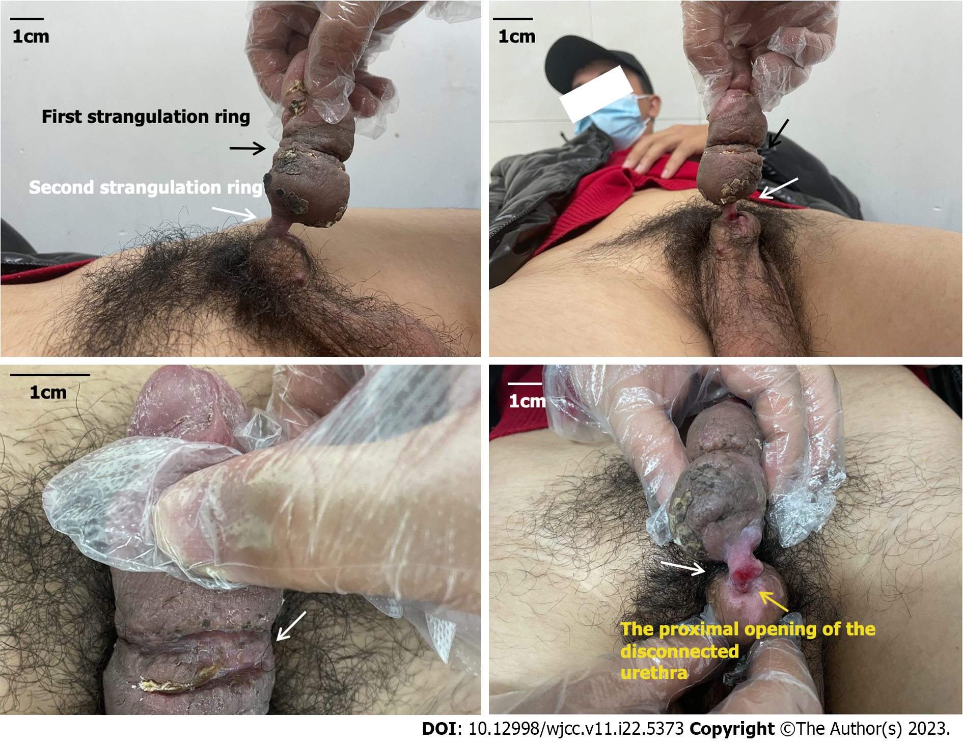
Figure 1 Preoperative physical examination.
The clinical presentation depicts a traumatic partial amputation on the proximal penile shaft with only a partial residual connection between the proximal and distal ends of the penis. The image shows the first tourniquet indicated by the black arrow, the second tourniquet indicated by the white arrow, and the proximal opening of the disconnected urethra indicated by the yellow arrow.
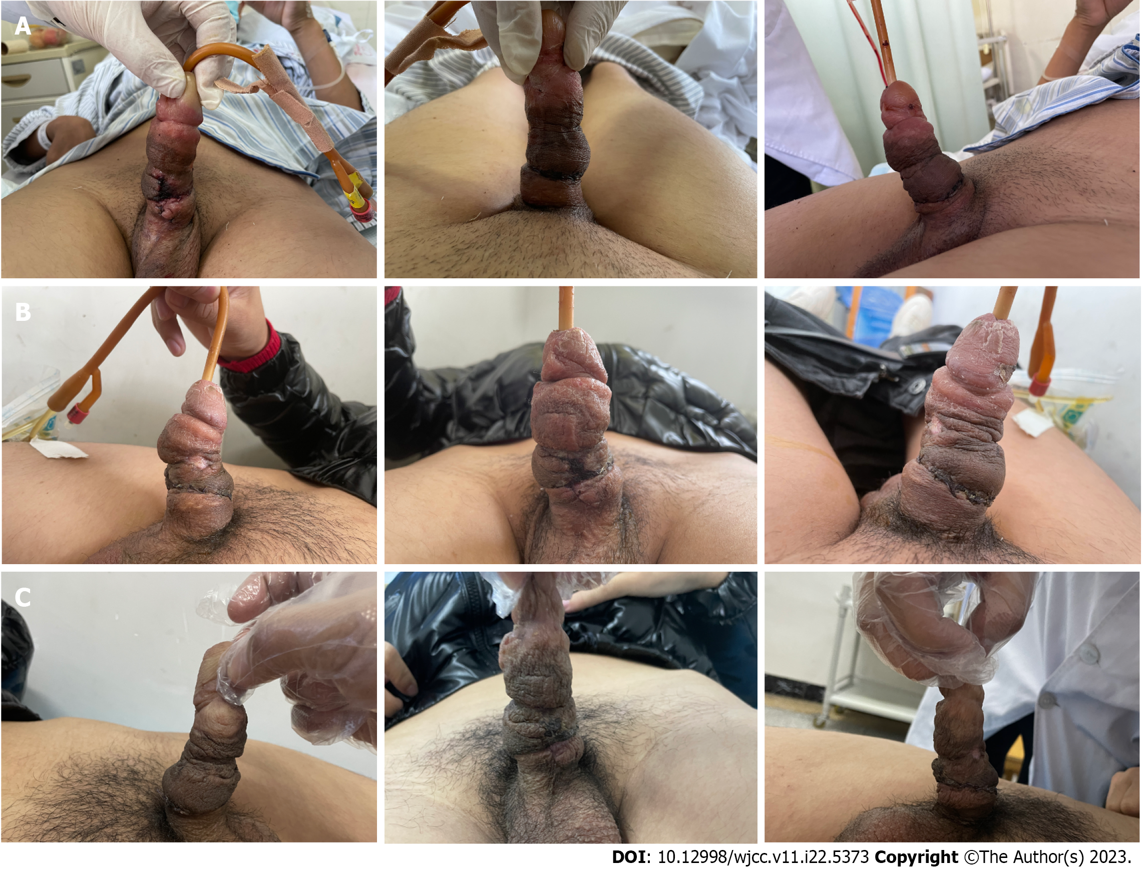
Figure 2 Postoperative follow-ups.
The image depicts the wound recovery status of patients at 3 d, 14 d, and 3 mo after surgical treatment. A: 3 d; B: 14 d; C: 3 mo.
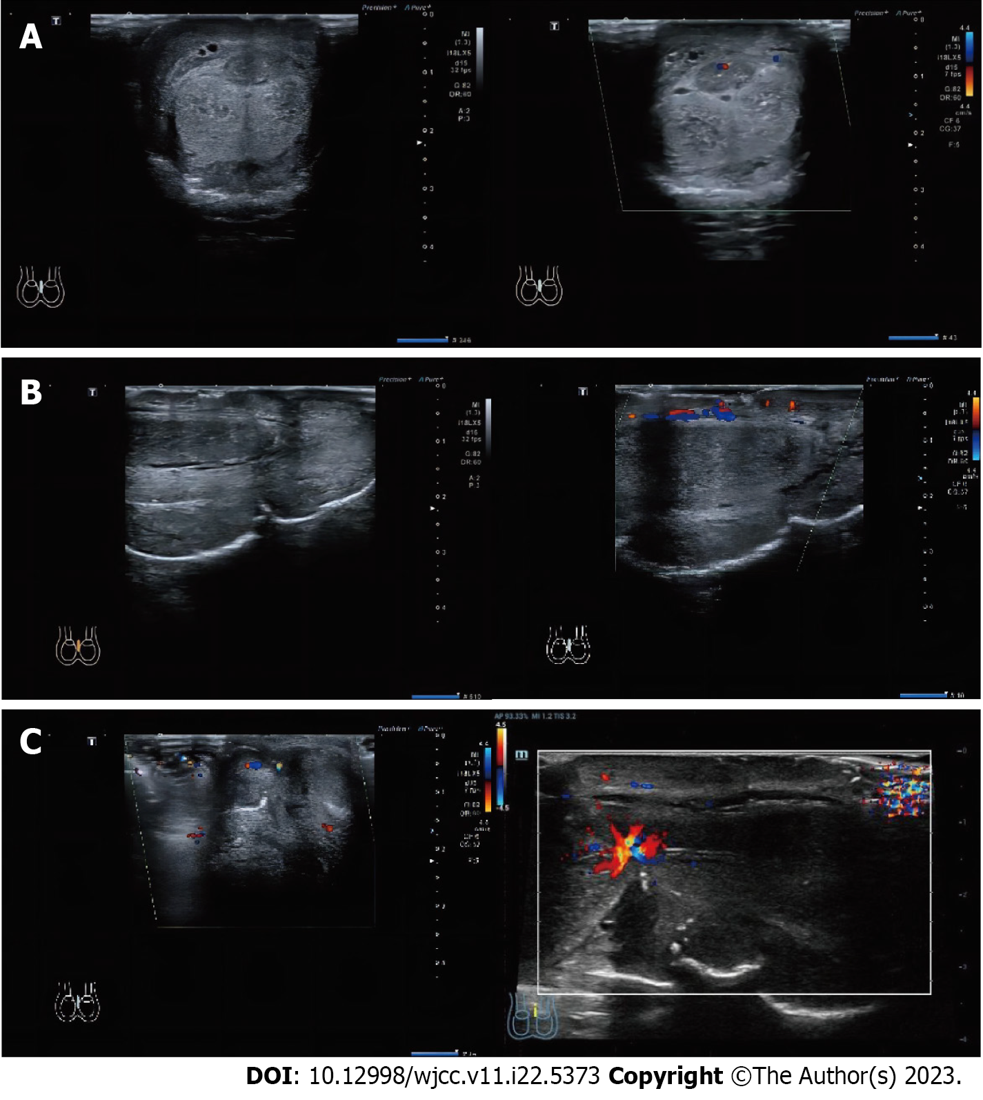
Figure 3 Preoperative Doppler ultrasonography.
The image depicts the color Doppler ultrasound display of the blood flow signal in the coronal and sagittal planes, as well as the strangulation site of the penis. A: Coronal plane; B: Vertical plane; C: Strangulation site.
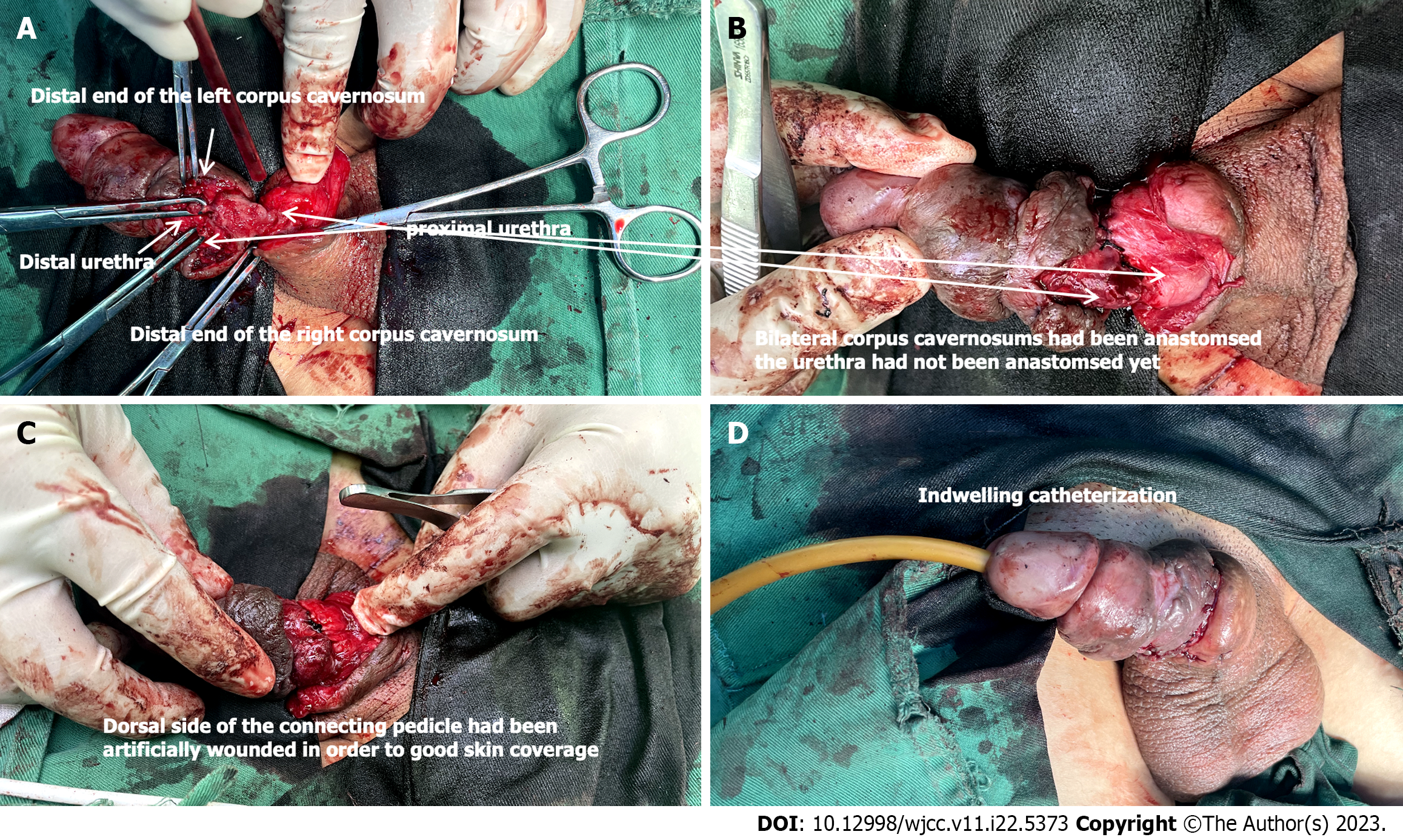
Figure 4 Surgery procedure.
Urethral anastomosis and reconstruction was performed. A: Adequately expose the pertinent anatomical structures of the surgical site; B: Approximate the severed corpus cavernosum of the penis; C: Implement local wound management to optimize skin coverage; D: Insertion of a urinary catheter.
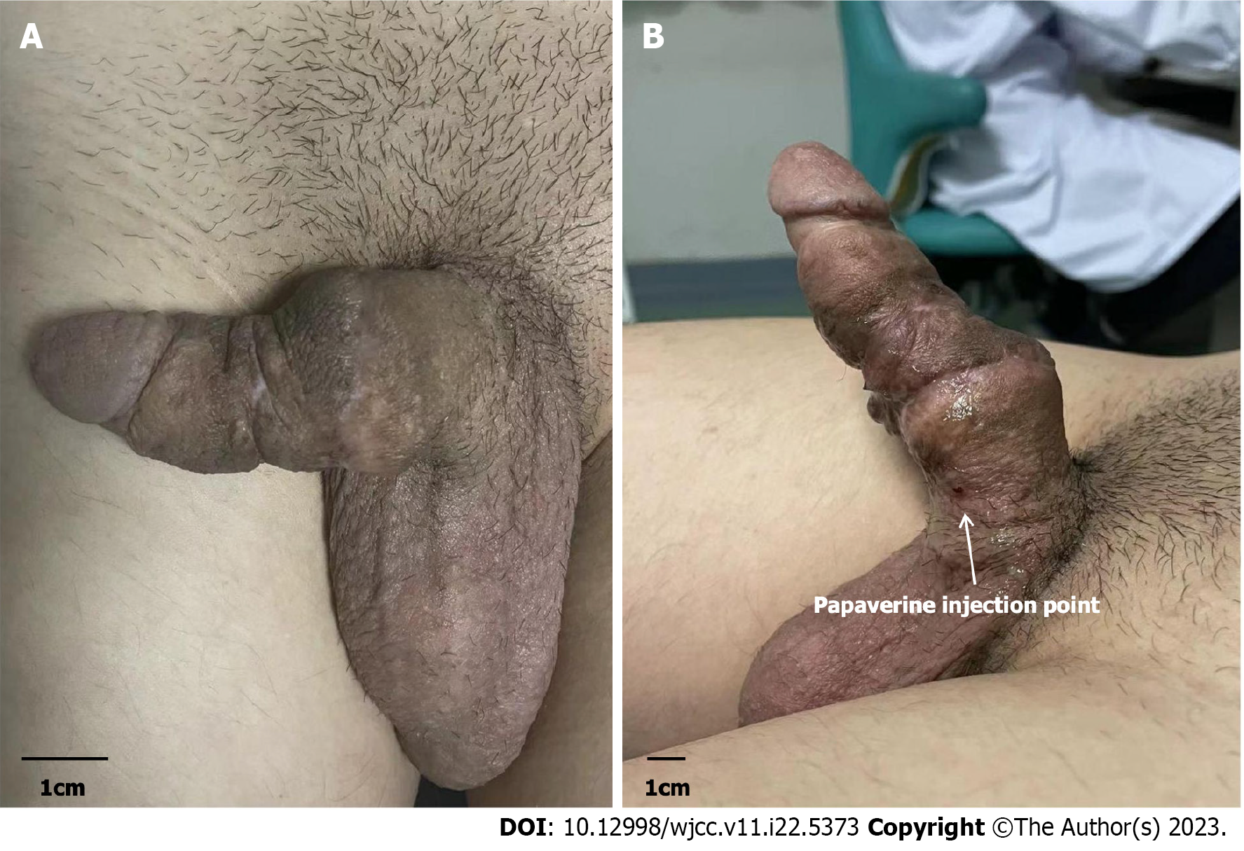
Figure 5 Postoperative pharmarco penile duplex color doppler ultrasonograghy 3 mo after surgery.
A: Natural state; B: 5 min after papaverine injection.
- Citation: Maimaitiming ABLT, Mulati YLSD, Apizi ART, Li XD. Self-strangulation induced penile partial amputation: A case report. World J Clin Cases 2023; 11(22): 5373-5381
- URL: https://www.wjgnet.com/2307-8960/full/v11/i22/5373.htm
- DOI: https://dx.doi.org/10.12998/wjcc.v11.i22.5373


