Copyright
©The Author(s) 2023.
World J Clin Cases. Aug 6, 2023; 11(22): 5365-5372
Published online Aug 6, 2023. doi: 10.12998/wjcc.v11.i22.5365
Published online Aug 6, 2023. doi: 10.12998/wjcc.v11.i22.5365
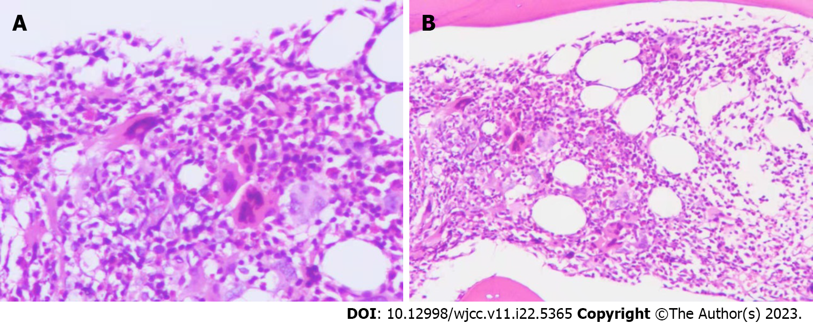
Figure 1 Bone marrow cell smear.
Oil microscopy, 1000 ×. Megakaryocytes are large and multi-lobulated.
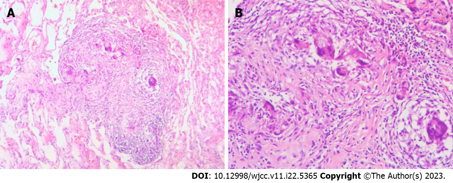
Figure 2 Bone marrow pathology images.
A: Large bone marrow megakaryocytes under high magnification (200 ×); B: Large bone marrow megakaryocytes under low magnification (100 ×).
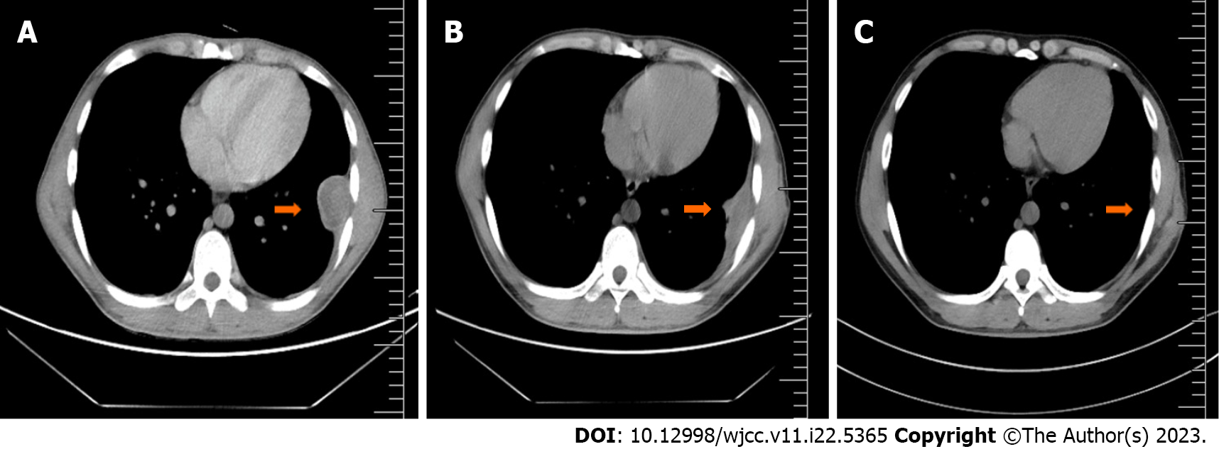
Figure 3 Pathological pictures of the chest wall mass.
A: Chest wall tuberculosis under low magnification (100 ×) showing epithelioidcells; B: Chest wall tuberculosis under high magnification (200 ×) showing epithelioid cells and granuloma.
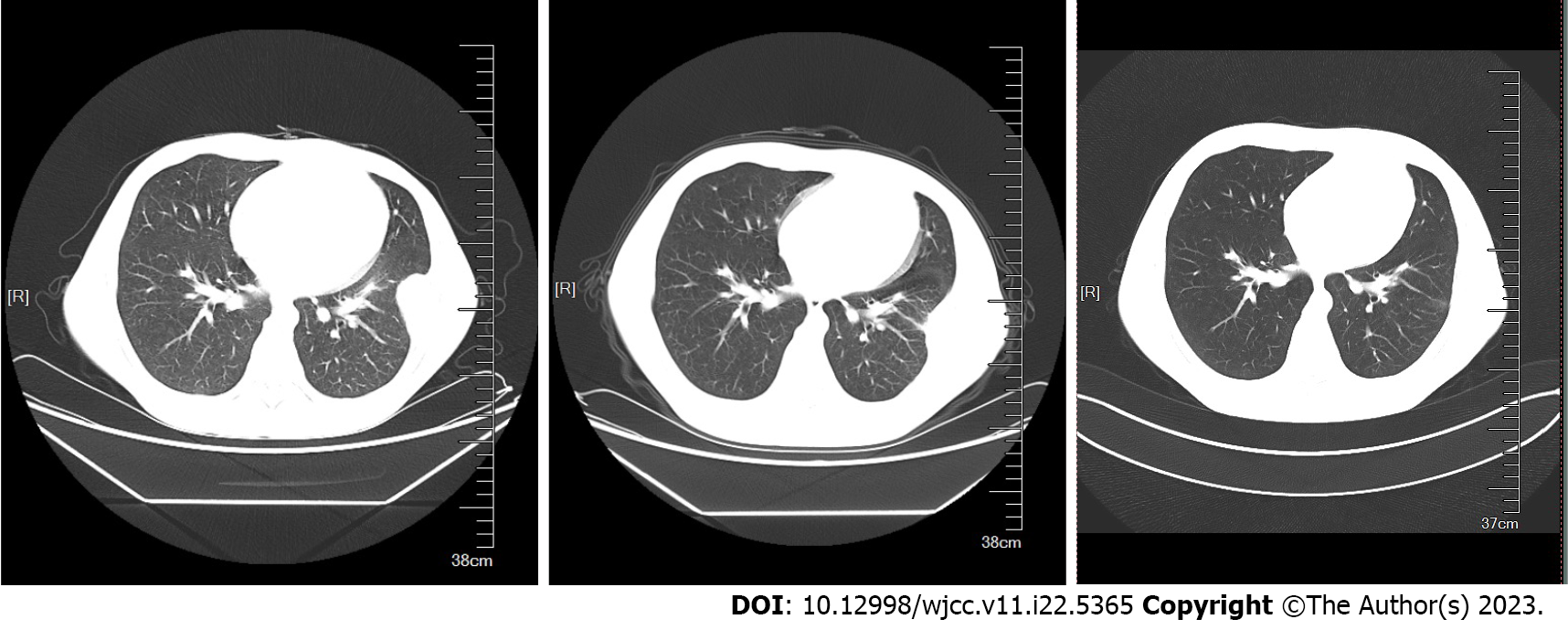
Figure 4 Chest computed tomography scan.
A: Taken July 27, 2017. A mound-shaped soft tissue density shadow with heterogeneous density can be seen under the left lower chest wall. Its size is about 43 mm × 34 mm × 65 mm, without obvious enhancement. Both the scan and enhancement computed tomography values were between 40-50 HU; B: Taken January 9, 2018. A small amount of fluid density shadow can be seen in the left thorax, partially wrapped; C: Taken June 11, 2021. The original mound-shaped soft tissue is no longer visible.
Figure 5 Chest wall computed tomography scan.
- Citation: Xu XY, Yang YB, Yuan J, Zhang XX, Kang L, Ma XS, Yang J. Individual with concurrent chest wall tuberculosis and triple-negative essential thrombocythemia: A case report. World J Clin Cases 2023; 11(22): 5365-5372
- URL: https://www.wjgnet.com/2307-8960/full/v11/i22/5365.htm
- DOI: https://dx.doi.org/10.12998/wjcc.v11.i22.5365


