Copyright
©The Author(s) 2023.
World J Clin Cases. Aug 6, 2023; 11(22): 5288-5295
Published online Aug 6, 2023. doi: 10.12998/wjcc.v11.i22.5288
Published online Aug 6, 2023. doi: 10.12998/wjcc.v11.i22.5288
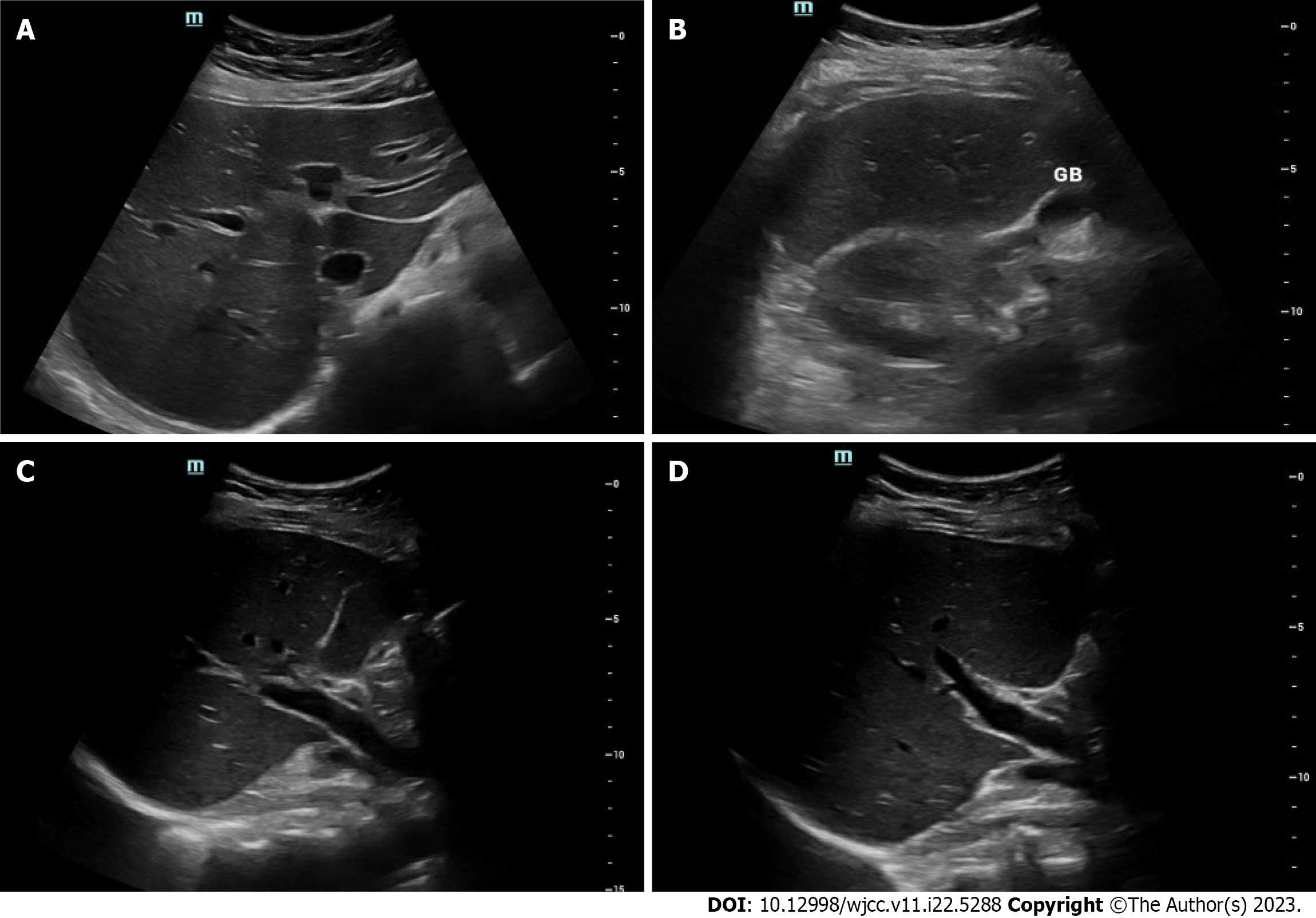
Figure 1 Ultrasound images of transverse section of the liver.
A: Left liver lobe; B: Right liver lobe; C: Main portal vein and branches; D: Hepatic artery.
Figure 2 Magnetic resonance cholangiopancreatography revealing no stones or ductal dilatation.
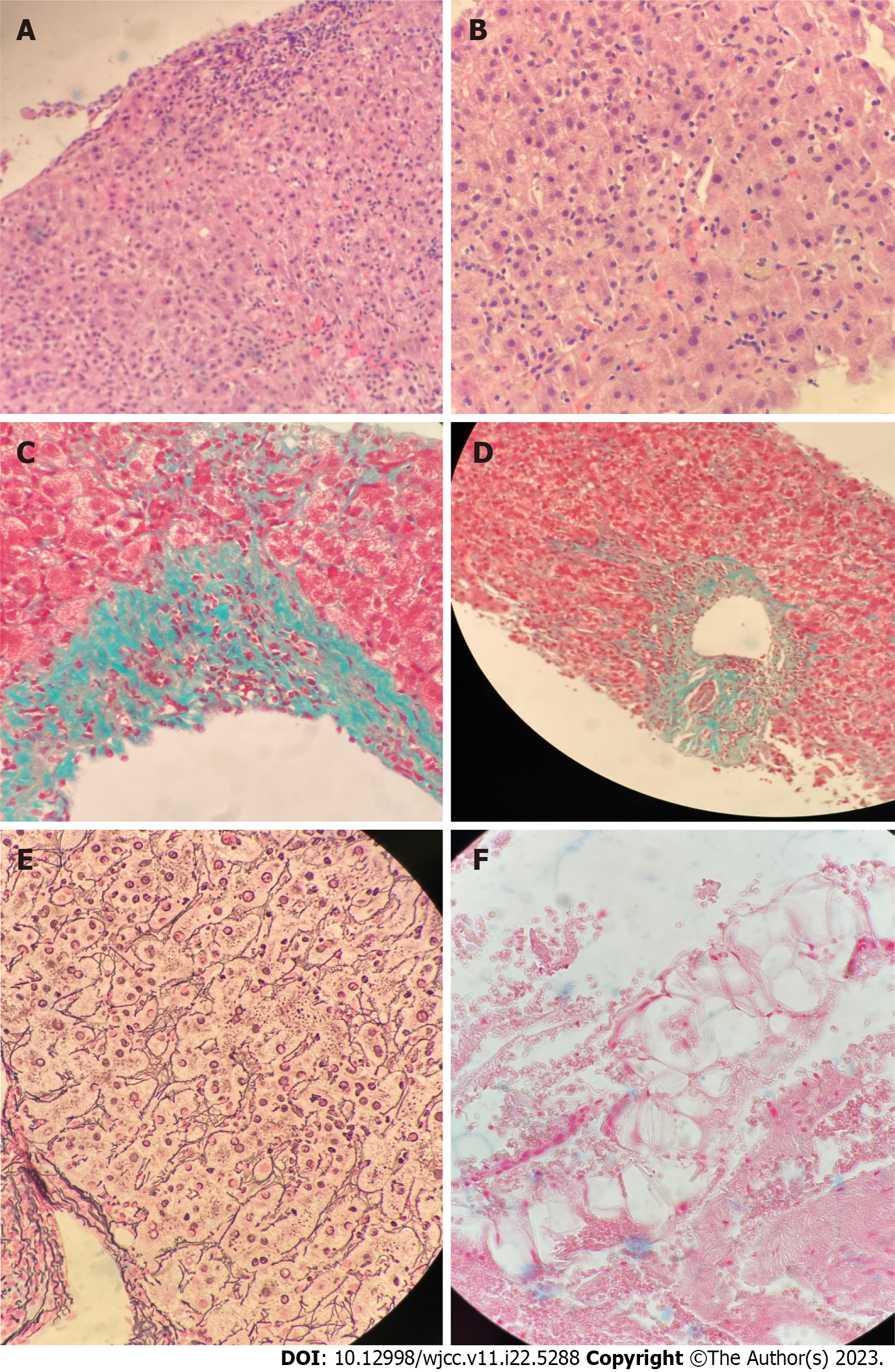
Figure 3 Histology images of liver biopsy with various stains.
A and B: H&E staining is negative for cholestasis, granulomas, and malignancy; C and D: Trichrome stain showing periportal fibrosis; E: Reticulin staining highlights lobular disarray; F: Iron stain is negative for siderosis.
- Citation: Dass L, Pacia AMM, Hamidi M. Acute hepatitis of unknown etiology in an adult female: A case report. World J Clin Cases 2023; 11(22): 5288-5295
- URL: https://www.wjgnet.com/2307-8960/full/v11/i22/5288.htm
- DOI: https://dx.doi.org/10.12998/wjcc.v11.i22.5288


