Copyright
©The Author(s) 2023.
World J Clin Cases. Jul 26, 2023; 11(21): 5179-5186
Published online Jul 26, 2023. doi: 10.12998/wjcc.v11.i21.5179
Published online Jul 26, 2023. doi: 10.12998/wjcc.v11.i21.5179
Figure 1 Ultrasound findings at 37 wk gestation showing foetal bilateral pleural effusion.
A: On coronal section; B: Sagittal section; C: Cross section.
Figure 2 High-throughput sequencing findings.
A: The precursor was consistent with maternal ADAMTS3 gene variants and that the father showed no abnormalities. The upper and middle panels represent the proband and maternal mutations, respectively. The lower panel displays the corresponding paternal sequencing results; B: The precursor was consistent with maternal FLT4 gene variants whereasthe father showed no abnormalities; C: The precursor was consistent with paternal AGT gene variants whereas the mother showed no abnormalities.
Figure 3 Gross anatomy of the pleuroperitoneal cavity.
A: Both lungs are poorly developed and the liver is abnormally enlarged; B: Autopsy findings revealed significant pleural effusion on the right side with poorly developed lungs.
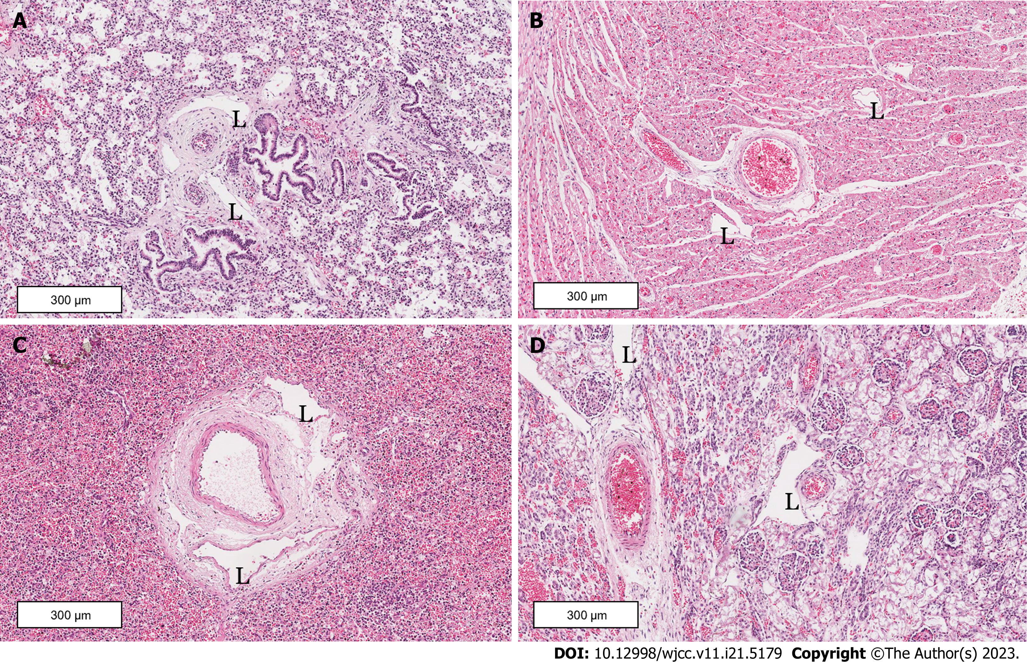
Figure 4 Autopsy findings.
A: Lung section illustrating vast abnormally dilated and tortuous lymphatic channels; B: Heart section; C: Liver section; D: Kidney section. L: Lymphatic channels.
- Citation: Liang ZW, Gao WL. ADAMTS3 and FLT4 gene mutations result in congenital lymphangiectasia in newborns: A case report. World J Clin Cases 2023; 11(21): 5179-5186
- URL: https://www.wjgnet.com/2307-8960/full/v11/i21/5179.htm
- DOI: https://dx.doi.org/10.12998/wjcc.v11.i21.5179


