Copyright
©The Author(s) 2023.
World J Clin Cases. Jul 26, 2023; 11(21): 5173-5178
Published online Jul 26, 2023. doi: 10.12998/wjcc.v11.i21.5173
Published online Jul 26, 2023. doi: 10.12998/wjcc.v11.i21.5173
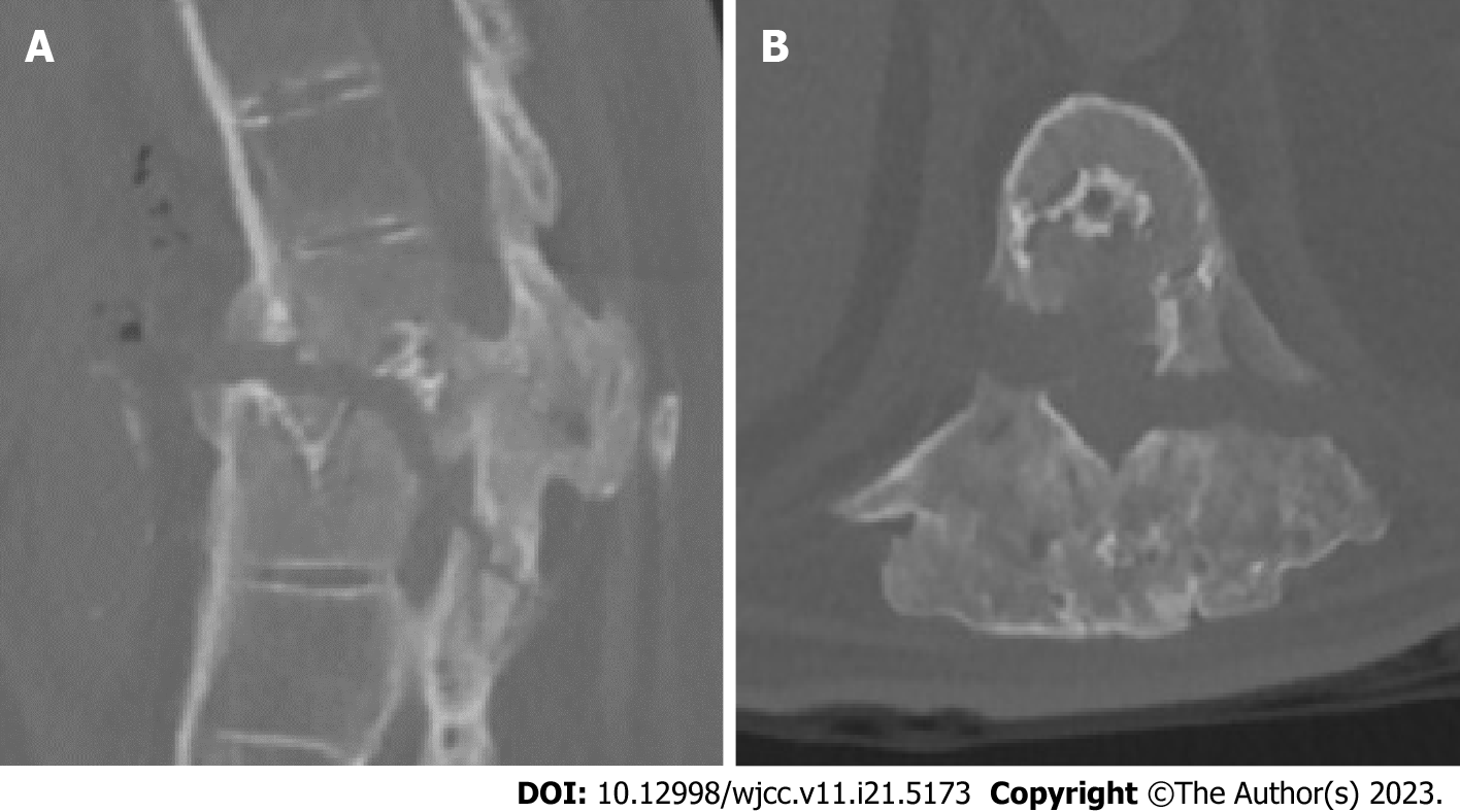
Figure 1 Computed tomography showed T10 and T11 fracture and spinal canal involvement.
Dislocation was also observed. A: Sagittal view; B: Axial view.
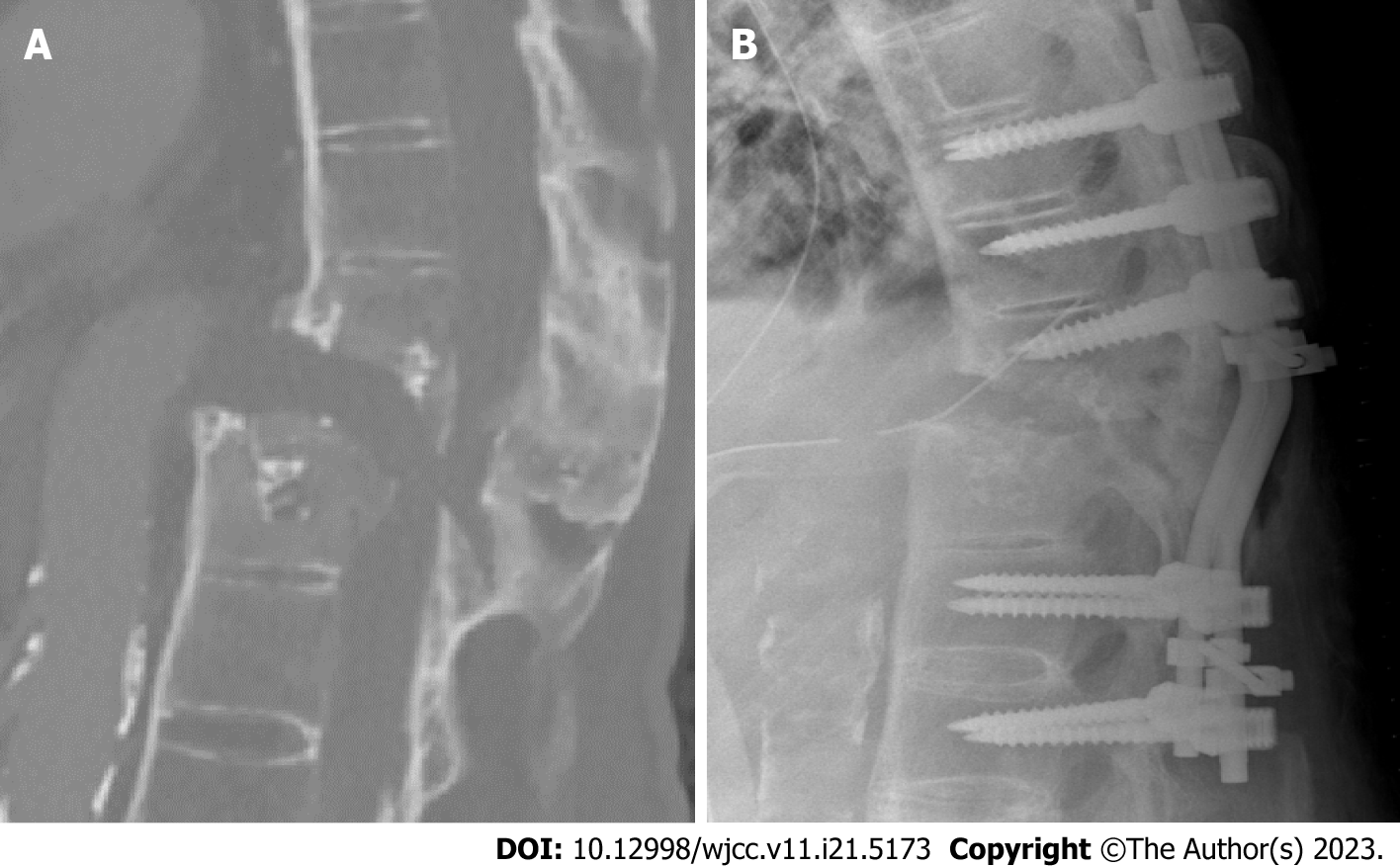
Figure 2 Imaging views of aggravated fracture and spinal cord stretching.
A: The progressive fracture dislocation with spinal cord stretching was revealed by repeat computed tomography (sagittal view) 3 d after video-assisted thoracoscopic surgery; B: Postoperative view by X ray (lateral view).
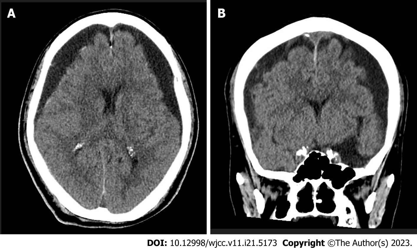
Figure 3 Bilateral hypodense subdural fluid accumulation with brain compression was revealed on brain computed tomography.
A: Axial view; B: Coronal view.
- Citation: Chen PH, Li CR, Gan CW, Yang TH, Chang CS, Chan FH. Rare combination of traumatic subarachnoid-pleural fistula and intracranial subdural hygromas: A case report. World J Clin Cases 2023; 11(21): 5173-5178
- URL: https://www.wjgnet.com/2307-8960/full/v11/i21/5173.htm
- DOI: https://dx.doi.org/10.12998/wjcc.v11.i21.5173


