Copyright
©The Author(s) 2023.
World J Clin Cases. Jul 26, 2023; 11(21): 5147-5159
Published online Jul 26, 2023. doi: 10.12998/wjcc.v11.i21.5147
Published online Jul 26, 2023. doi: 10.12998/wjcc.v11.i21.5147
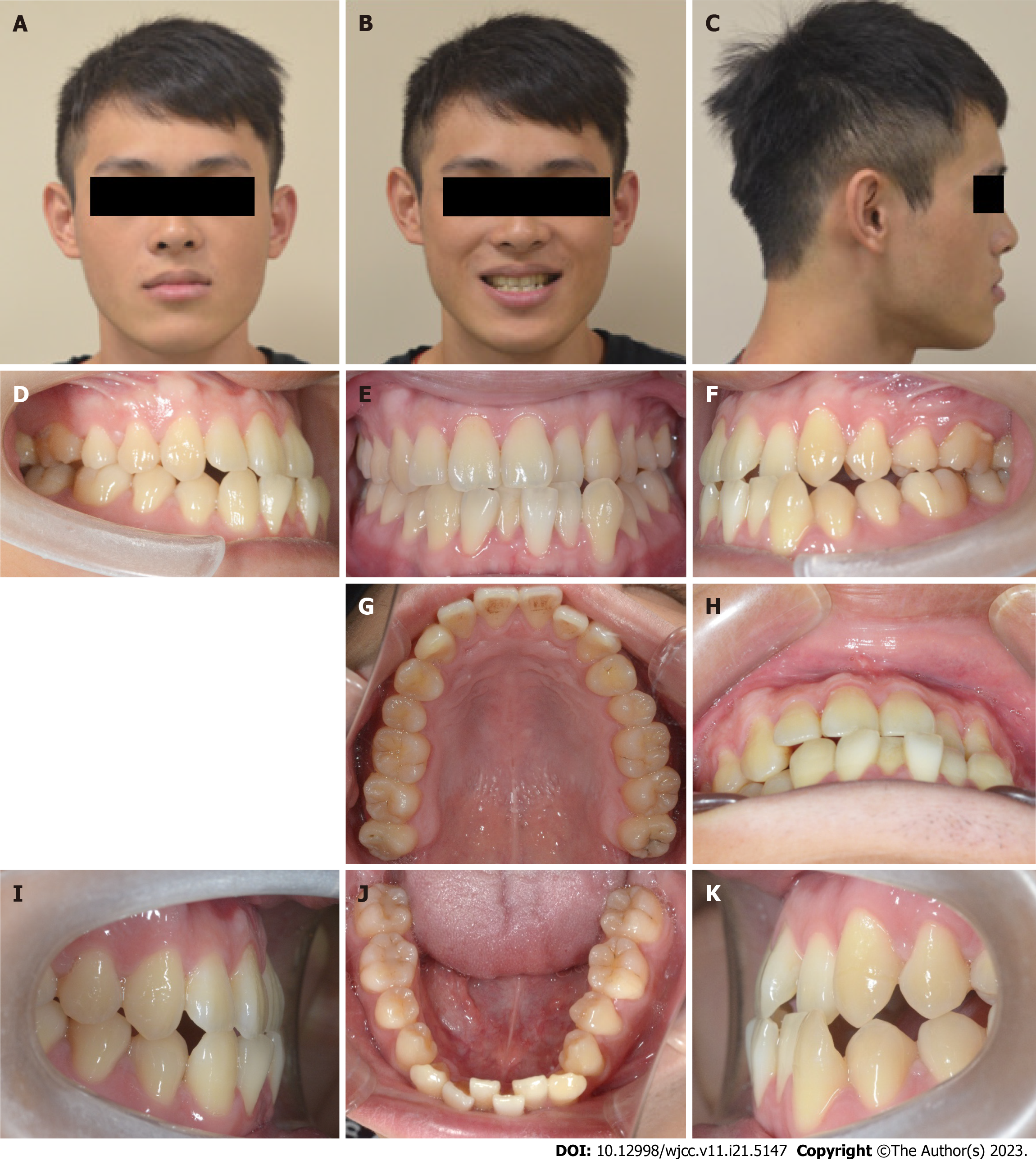
Figure 1 Pretreatment facial and intraoral photographs.
A: Patient's facial photograph, frontal view at rest; B: Patient's facial photograph, frontal view smiling; C: Patient's facial photograph, lateral view at rest; D: Right-side occlusal view; E: Frontal view; F: Left-side occlusal view; G: Maxillary arch; H: Overbite and overjet view; I: Right-side overbite and overjet view; J: Mandibular arch; K: Left-side overbite and overjet view.
Figure 2 Pretreatment dental casts.
A-C: View showing occlusal relationship; D and E: View showing maxillary and mandibular arch.
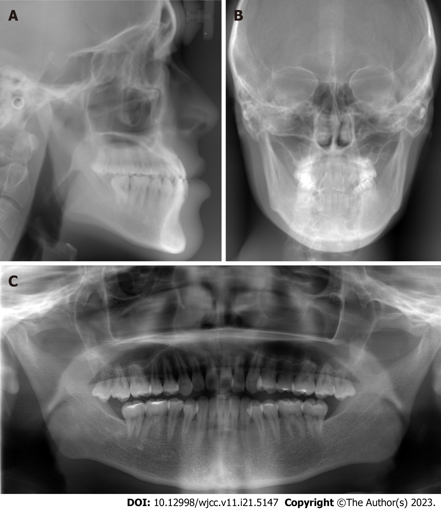
Figure 3 Pretreatment X-ray images.
A: Lateral cephalogram; B: Frontal cephalogram; C: Panoramic radiograph.
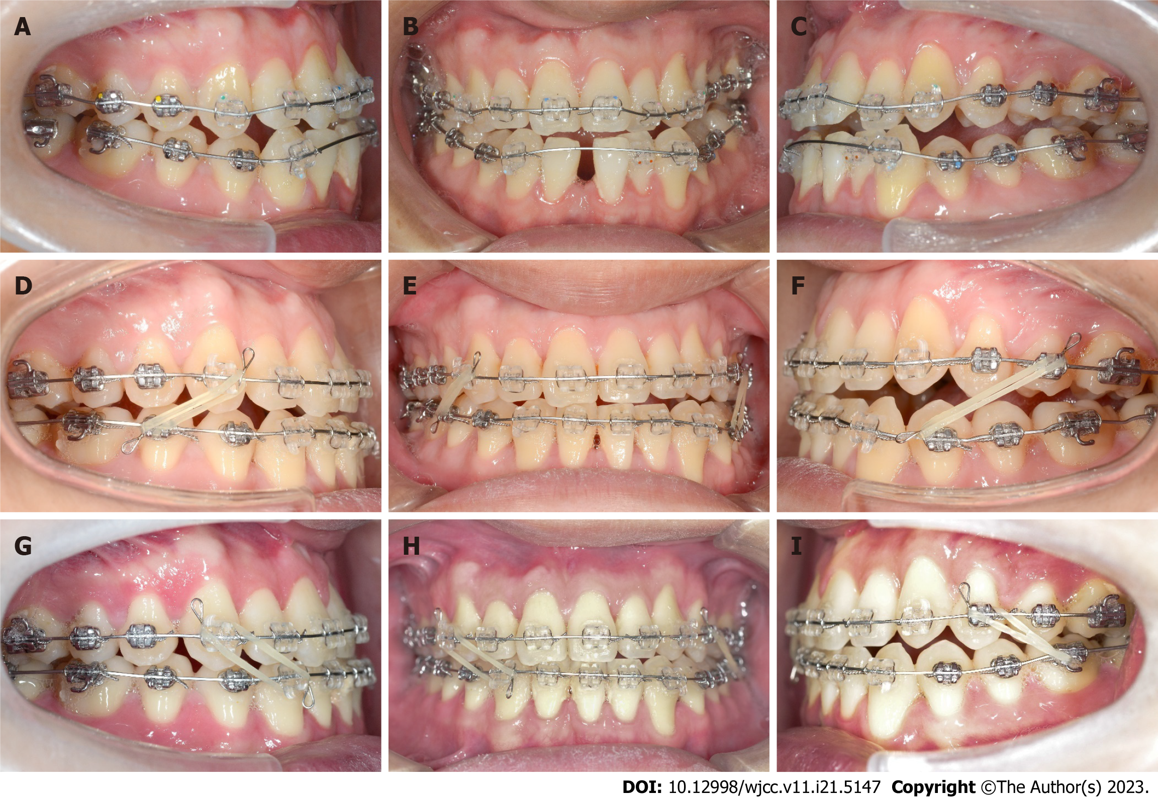
Figure 4 Tooth movement changes during treatment.
A-C: Intraoral photographs during initial alignment; D-F: Intraoral photographs after midline correction; G-I: Intraoral photographs during correction of facial asymmetry.
Figure 5 Mechanics to intrude the canine (not-in-slot).
Figure 6 Posttreatment facial and intraoral photographs.
A: Patient's facial photograph, frontal view at rest; B: Patient's facial photograph, frontal view smiling; C: Patient's facial photograph, lateral view at rest; D: Maxillary arch; E: Overbite and overjet view; F: Right-side occlusal view; G: Frontal view; H: Left-side occlusal view; I: Right-side overbite and overjet view; J: Mandibular arch; K: Left-side overbite and overjet view.
Figure 7 Posttreatment dental casts.
A-C: View showing occlusal relationship; D and E: View showing maxillary and mandibular arch.
Figure 8 Posttreatment X-ray images.
A: Lateral cephalogram; B: Frontal cephalogram; C: Panoramic radiograph.
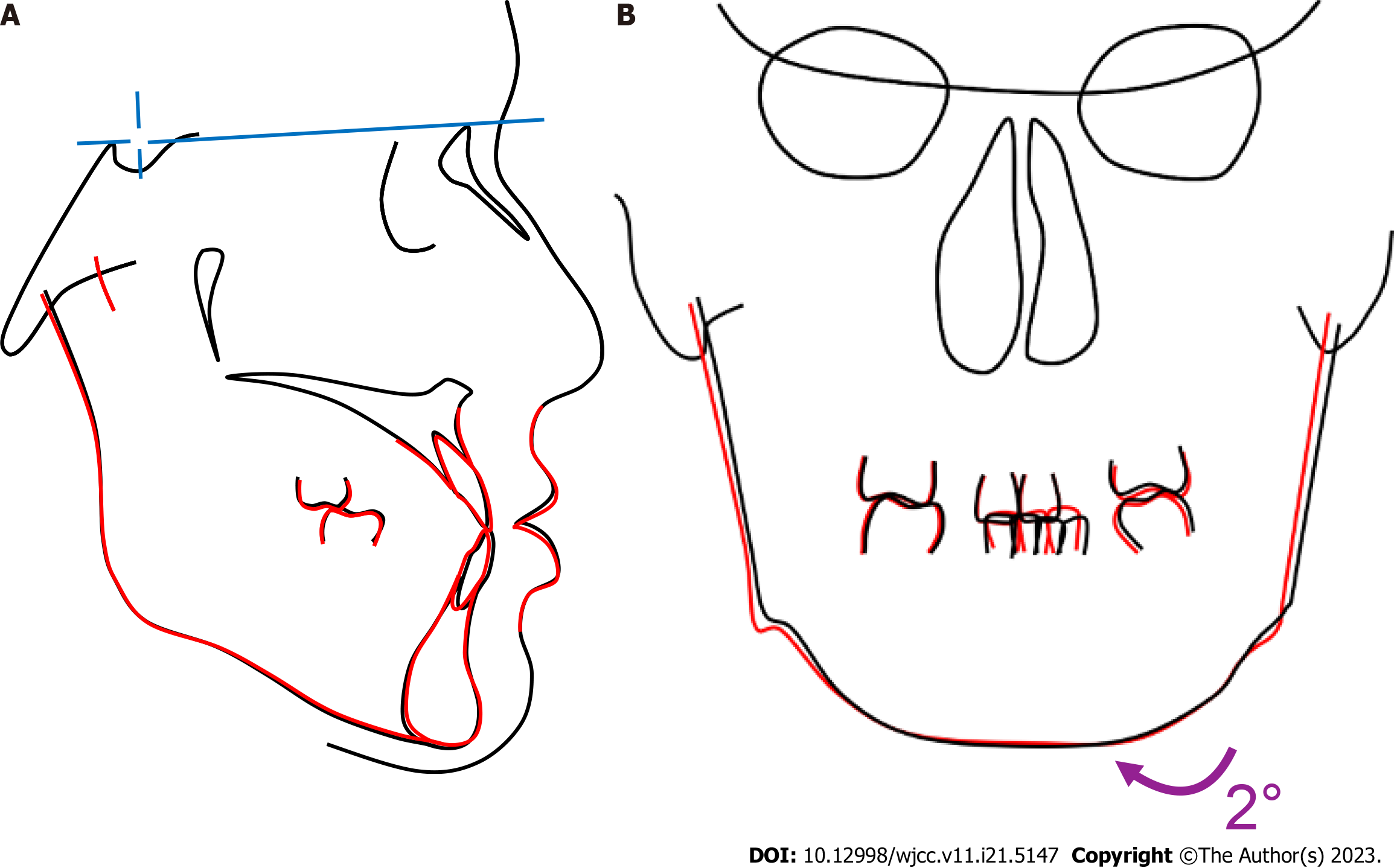
Figure 9 Superimposition images: Pretreatment (black) and posttreatment (red).
A: Lateral cephalograms; B: Frontal cephalograms.
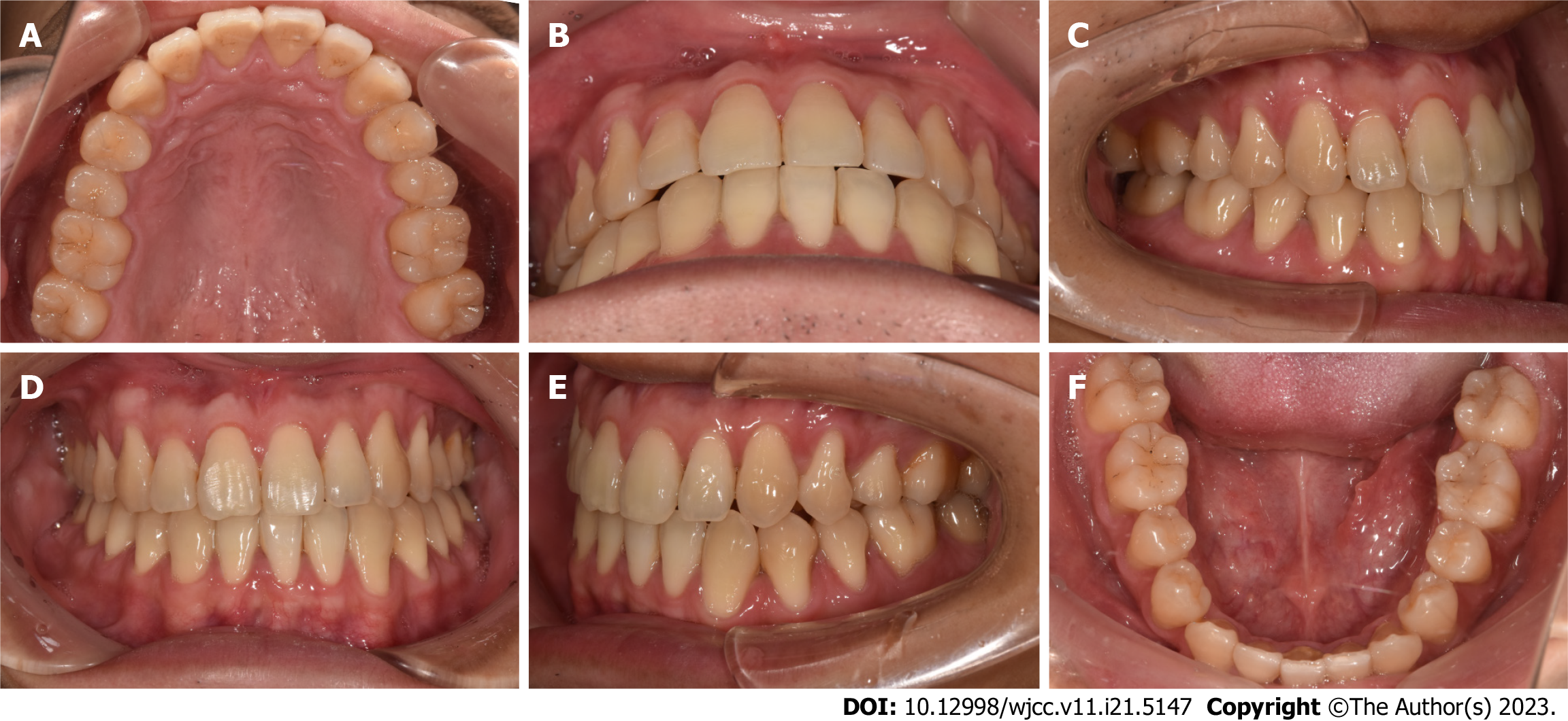
Figure 10 One-year follow-up intraoral photographs.
A: Maxillary arch view; B: Overbite and overjet view; C: Right-side occlusal view; D: Frontal view; E: Left-side occlusal view; F: Mandibular arch view.
Figure 11 Two-years follow-up intraoral photographs.
A: Maxillary arch view; B: Overbite and overjet view; C: Right-side occlusal view; D: Frontal view; E: Left-side occlusal view; F: Mandibular arch.
Figure 12 Three-years follow-up intraoral photographs.
A: Maxillary arch view; B: Overbite and overjet view; C: Right-side occlusal view; D: Frontal view; E: Left-side occlusal view; F: Mandibular arch.
- Citation: Huang CY, Chen YH, Lin CC, Yu JH. Improved super-elastic Ti-Ni alloy wire for treating adult skeletal class III with facial asymmetry: A case report. World J Clin Cases 2023; 11(21): 5147-5159
- URL: https://www.wjgnet.com/2307-8960/full/v11/i21/5147.htm
- DOI: https://dx.doi.org/10.12998/wjcc.v11.i21.5147


