Copyright
©The Author(s) 2023.
World J Clin Cases. Jul 26, 2023; 11(21): 5136-5146
Published online Jul 26, 2023. doi: 10.12998/wjcc.v11.i21.5136
Published online Jul 26, 2023. doi: 10.12998/wjcc.v11.i21.5136
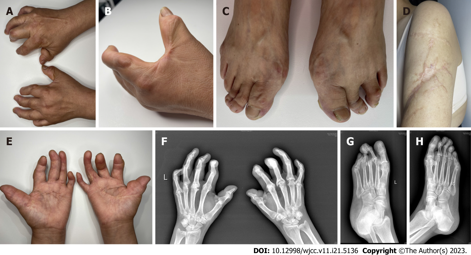
Figure 1 Pathological changes of skin and joints of limbs in patients with fibroblast rheumatism.
A and B: The patient's fingers showing swelling with flexion contractures and the presence of numerous smooth, firmly, freely mobile nodules on the extensor surface of the palms, upper arms, MCP joints, and PIP joints; C: Notable contracture and multiple reddish firm nodules observed in the MTP and DIP joints; D: Scar connective tissue prominently located in the lateral margin of both upper arms; E: Joint flexion contractures evident in the patient's hands, particularly affecting all fingers, the thumb, ring, and little fingers, resulting in a 'main en griffe' (claw hand) appearance; F: Hand rand radiography revealing knuckle and interphalangeal joint flexion in in both palms, narrow joint spaces, partial fusion, and swan-neck changes in some DIP joints; G and H: Foot radiography showing a missing third toe bone in the left foot, narrow joint gaps between the two feet, flexion of the metatarsal toe joints, and semi-dislocation of the left foot's second metatarsophalangeal joint.
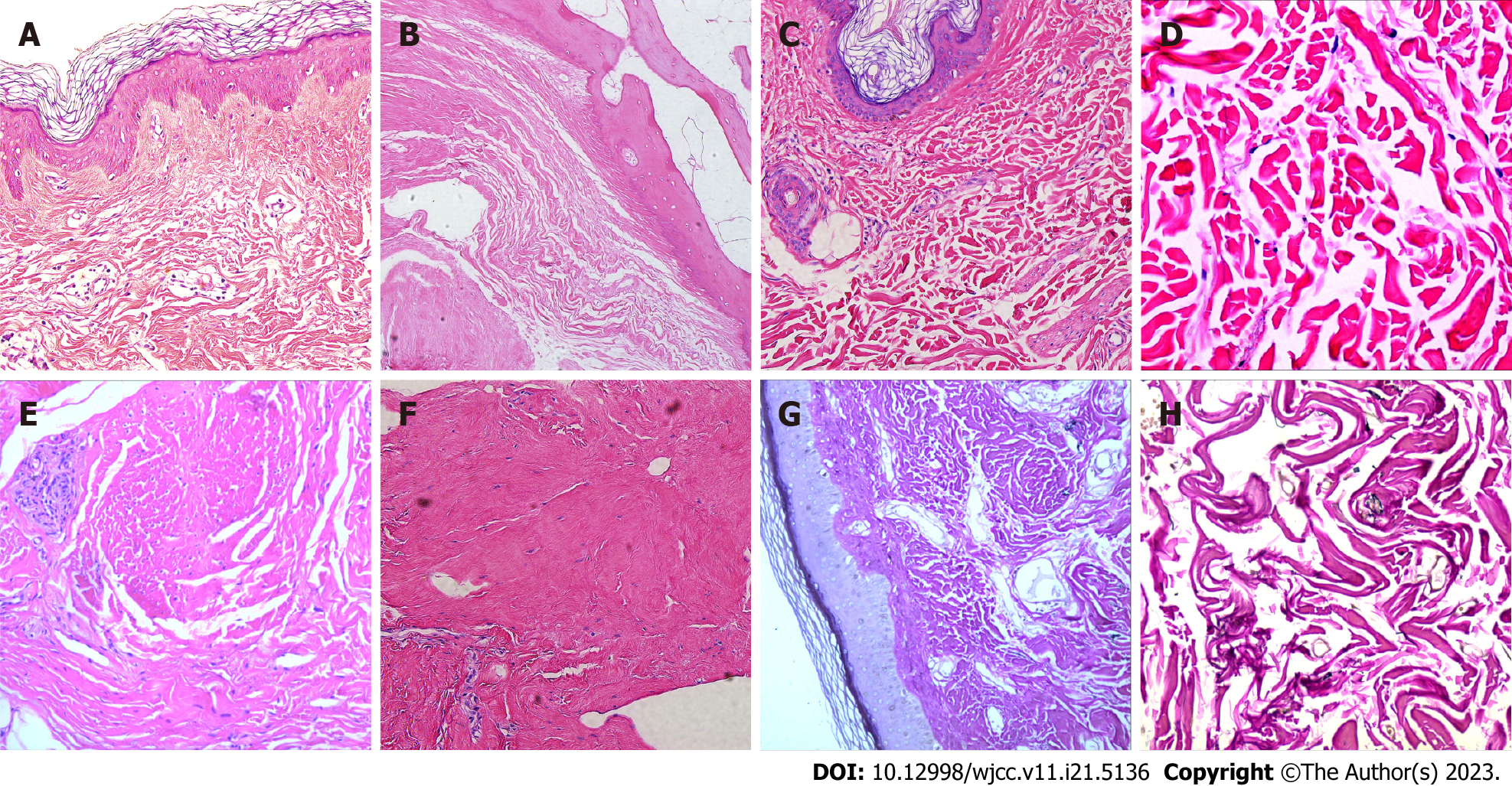
Figure 2 Typical pathological images of skin lesions in patients with fibroblast rheumatism.
A: Skin biopsy from the finger revealing dermal fibrous hyperplasia, elastic fiber disorder, skin hyperkeratosis, and irregular thickening of the spinous layer; B: Haematoxylin and eosin staining of left foot articular cartilage displaying central cartilage degeneration and necrosis with calcification, along with dermal collagen fiber hyperplasia and spindle cell proliferation; C and D: Histological examination of upper arm scar tissue and synovium, showing collagen fiber hyperplasia, elastic fiber disorder, and spindle cell proliferation; E and F: Palmar aponeurosis biopsy from the right arch demonstrating fibrous hyperplasia with localized spindle cell proliferation (Haematoxylin and eosin, original magnifications: (A: ×200; B: ×200; C: ×200; D: ×400; E: ×40; F: ×200); G and H: Elastica van Gieson staining revealing reduced elastic fibers in the dermis (G: ×200; H: ×400).
Figure 3 Immunohistochemical histopathologic images of skin lesions in patients with fibroblast rheumatism.
A: Positive staining for α-smooth muscle actin (α-SMA) (×200); B: Vimentin (×200) in the spindle cells; C: As well as positive staining for CD34 (×200); D: Negative staining observed for CD68 (×200), E: CD163 (×200); F: S100 (×200).
Figure 4 Preferred reporting items of Systematic Reviews and Meta-Analysis flow chart for the systematic review screening process.
- Citation: Guo H, Liang Q, Dong C, Zhang Q, Gu ZF. Systematic review of fibroblastic rheumatism: A case report. World J Clin Cases 2023; 11(21): 5136-5146
- URL: https://www.wjgnet.com/2307-8960/full/v11/i21/5136.htm
- DOI: https://dx.doi.org/10.12998/wjcc.v11.i21.5136


