Copyright
©The Author(s) 2023.
World J Clin Cases. Jul 26, 2023; 11(21): 5129-5135
Published online Jul 26, 2023. doi: 10.12998/wjcc.v11.i21.5129
Published online Jul 26, 2023. doi: 10.12998/wjcc.v11.i21.5129
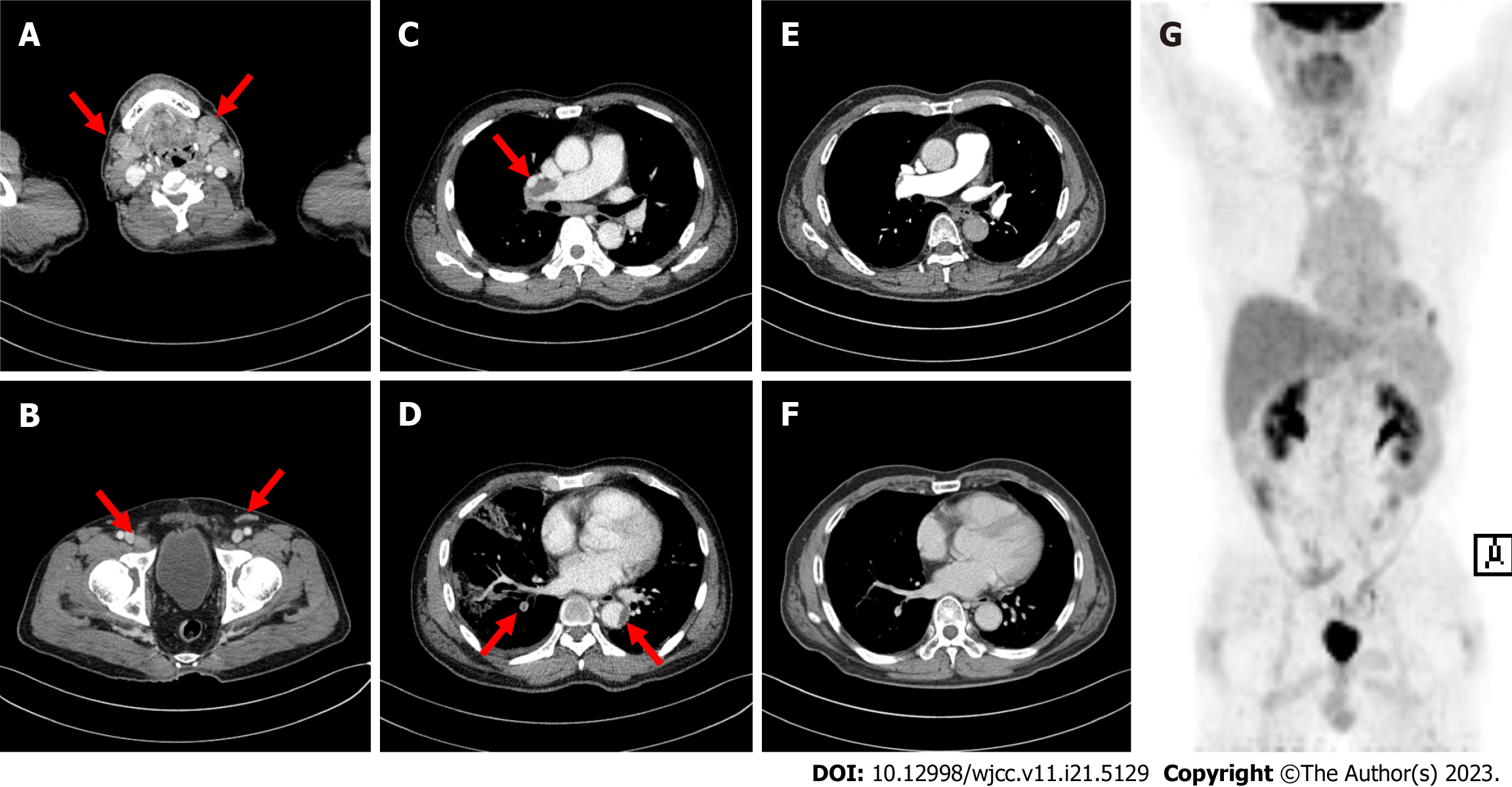
Figure 1 Imaging examination of the T-lymphoblastic lymphoma and pulmonary thromboembolism.
A-D: Multiple enlarged lymph nodes (A and B) and thromboembolism (C and D) visible on computed tomography (CT); E-G: CT (E and F) and positron emission tomography/CT (G) confirm the complete resolution of thromboembolism and complete response of T-lymphoblastic lymphoma, respectively.
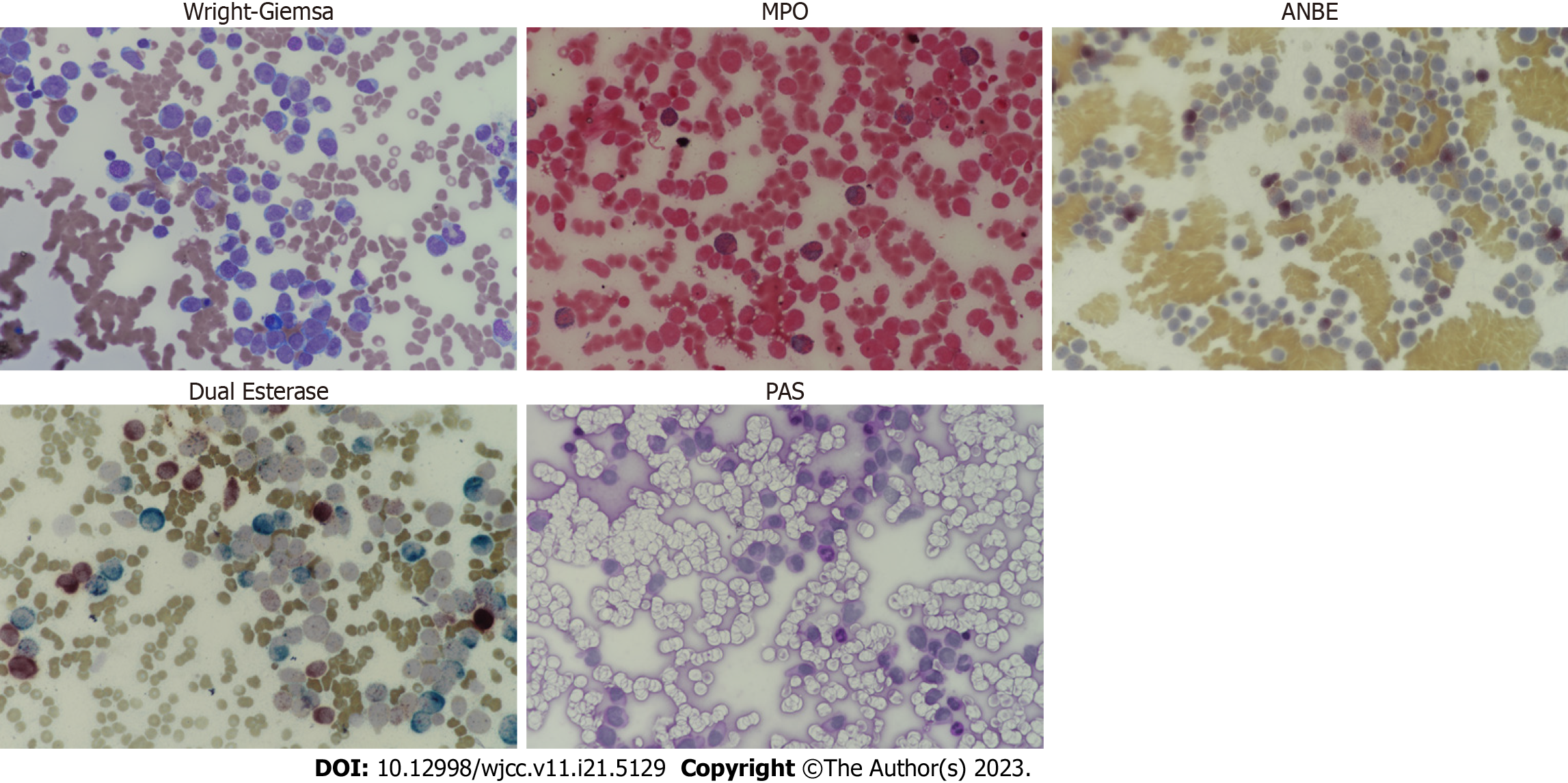
Figure 2 Bone marrow findings.
87% of all nucleated cells in the bone marrow are leukemic blasts. Cytochemically, the leukemic cells are positive for myeloperoxidase, myeloid marker and alpha-naphthyl butyrate esterase, monocyte marker. Dual esterase stain including alpha-naphthyl acetate esterase, another nonspecific esterase and chloroacetate esterase reveals that leukemic cells are derived from monocytes. While leukemic cells are negative for Periodic acid-Schiff stain. MPO: myeloperoxidase; ANBE: alpha-naphthyl butyrate esterase; PAS: Periodic acid-Schiff.
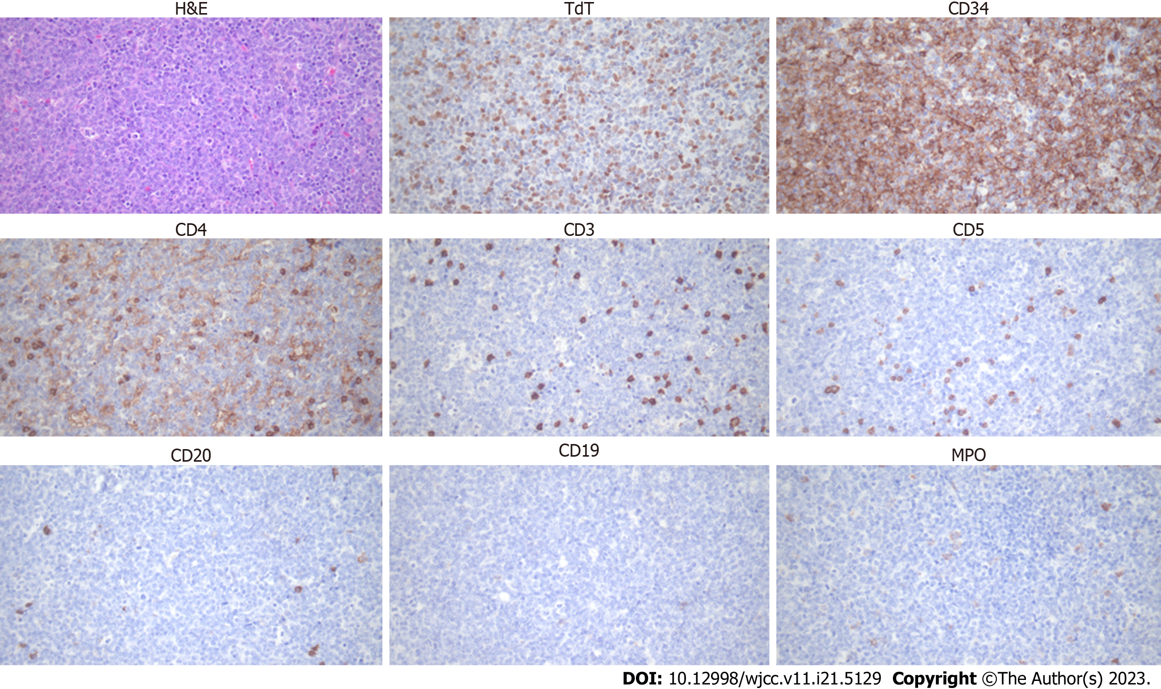
Figure 3 Histologic findings.
Tumor cells appear as a sheet and are medium-to-large-sized. Immunohistochemically, the tumor cells are moderately positive for terminal deoxynucleotidyl transferase, diffusely positive for cluster of differentiation (CD)34, weakly positive for CD4, and negative for CD3, CD5, CD20, CD19, and myeloperoxidase. H&E: Hematoxylin and eosin; TdT: Terminal deoxynucleotidyl transferase; CD: Cluster of differentiation; MPO: Myeloperoxidase.
- Citation: Jeon SY, Lee NR, Cha S, Yhim HY, Kwak JY, Jang KY, Kim N, Cho YG, Lee CH. Acute myelomonocytic leukemia and T-lymphoblastic lymphoma as simultaneous bilineage hematologic malignancy treated with decitabine: A case report. World J Clin Cases 2023; 11(21): 5129-5135
- URL: https://www.wjgnet.com/2307-8960/full/v11/i21/5129.htm
- DOI: https://dx.doi.org/10.12998/wjcc.v11.i21.5129


