Copyright
©The Author(s) 2023.
World J Clin Cases. Jul 26, 2023; 11(21): 5115-5121
Published online Jul 26, 2023. doi: 10.12998/wjcc.v11.i21.5115
Published online Jul 26, 2023. doi: 10.12998/wjcc.v11.i21.5115
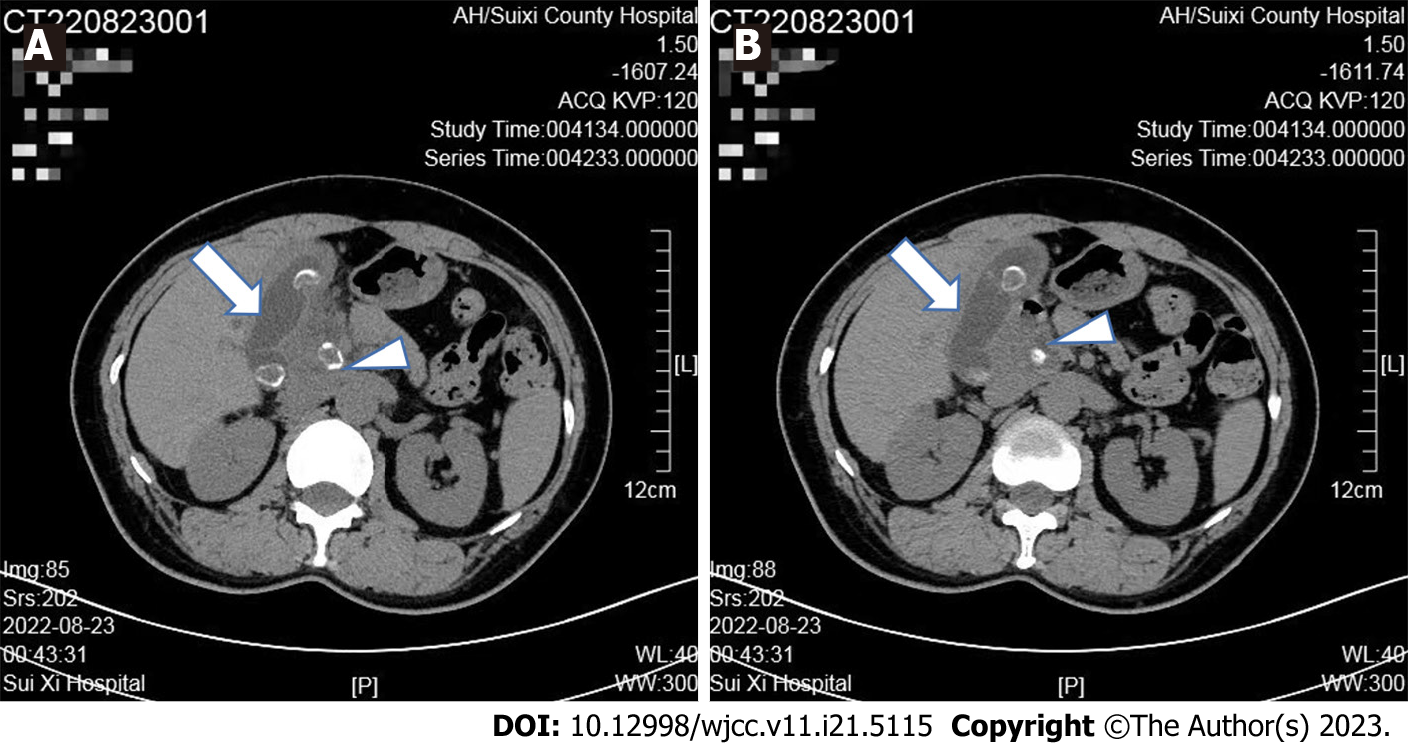
Figure 1 Abdominal computed tomography showed.
A: Stones at the junction of the common hepatic duct (white arrowhead) and gall bladder (white arrow); B: Multiple stones in the gallbladder, leading to cholecystitis (white arrow).
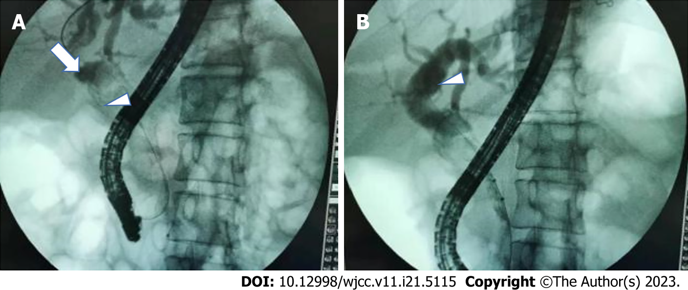
Figure 2 Endoscopic retrograde cholangiopancreatography radiography showed.
A: The bile ducts were visualized. The upper segment of the common bile duct was dilated (white arrow). In addition, an oval filling defect shadow existed at the junction of the cystic duct and common hepatic duct and moved (white arrowhead), and the junction of the cystic duct was narrowed; B: The common hepatic duct and intrahepatic bile duct were dendritically dilated, with multiple filling defect shadows in the gallbladder (white arrowhead).
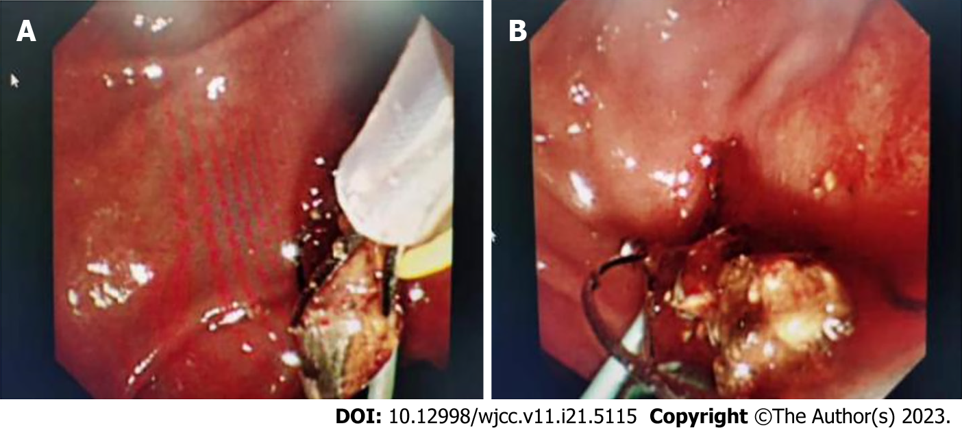
Figure 3 Electrohydraulic lithotripsy under the direct view of SpyGlass.
A: The stone impacted in the cystic duct partially went out into the lumen of the common bile duct under the direct view of the SpyGlass choledochoscopy; B: Electrohydraulic lithotripsy was performed under direct view of the SpyGlass.
Figure 4 Calculus was removed under duodenoscope.
A: Calculus was being removed with the Dormia basket out of the papilla of the duodenum; B: Calculus was removed completely.
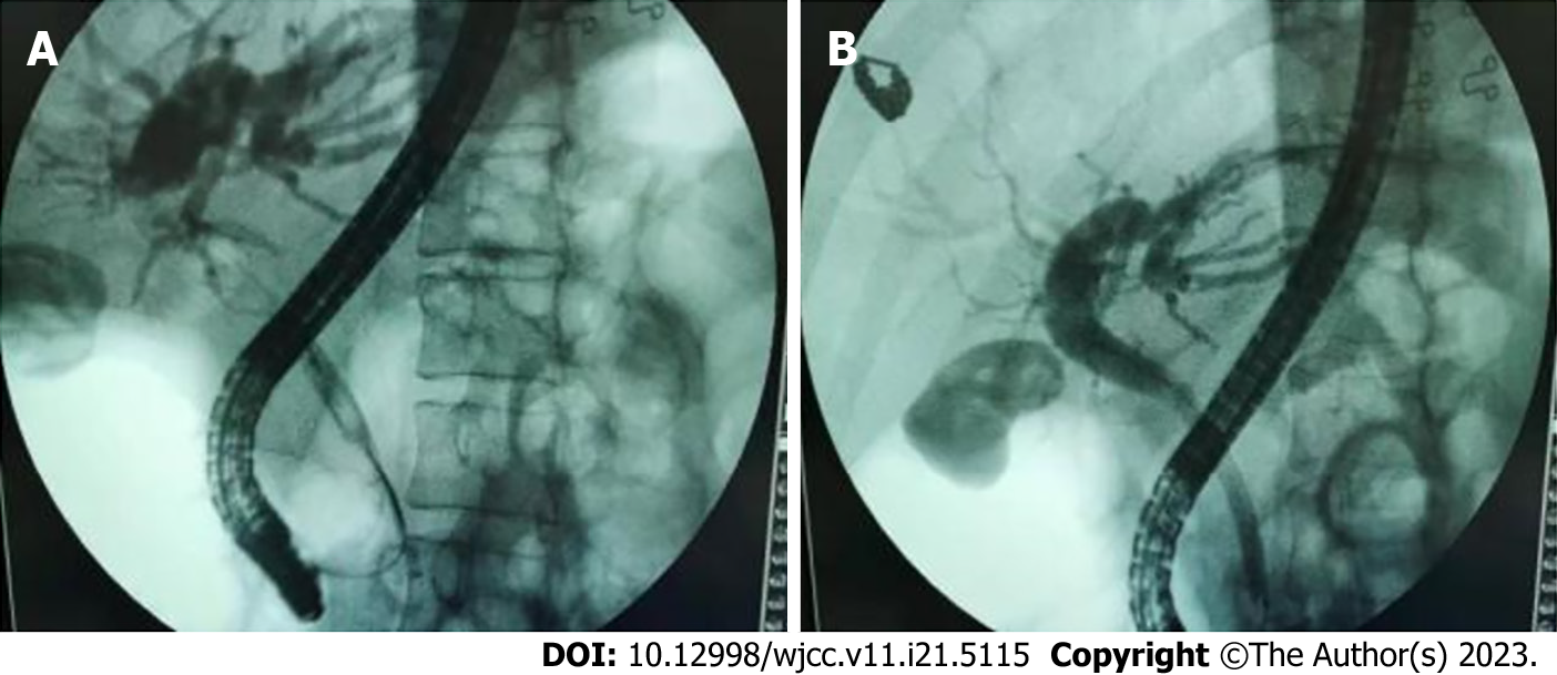
Figure 5 Postoperative radiography shows disappearance of the filling defect shadow.
A: After lithotripsy, the stones were repeatedly removed from the common bile duct with the Dormia basket and retrieval balloon; B: The filling defect shadow disappeared on the control endoscopic retrograde cholangio
- Citation: Liang SN, Jia GF, Wu LY, Wang JZ, Fang Z, Wang SH. Type I Mirizzi syndrome treated by electrohydraulic lithotripsy under the direct view of SpyGlass: A case report. World J Clin Cases 2023; 11(21): 5115-5121
- URL: https://www.wjgnet.com/2307-8960/full/v11/i21/5115.htm
- DOI: https://dx.doi.org/10.12998/wjcc.v11.i21.5115


